Explore
 Validate
 Learn
Learn Flow cytometry
Antibody data
- Antibody Data
- Antigen structure
- References [1]
- Comments [0]
- Validations
- Flow cytometry [1]
Submit
Validation data
Reference
Comment
Report error
- Product number
- 50-9046-41 - Provider product page

- Provider
- Invitrogen Antibodies
- Product name
- Anti-Phospho-ATM (Ser1981) Monoclonal Antibody (10H11.E12), eFluor 660, eBioscience™
- Antibody type
- Monoclonal
- Antigen
- Other
- Description
- Description: This 10H11.E12 monoclonal antibody recognizes the human, mouse, and rat ataxia-telangiectasia (ATM) protein when phosphorylated on serine 1981. ATM belongs to the family of PI3 kinases and functions by coordinating cell cycle arrest and initiation of DNA repair through the phosphorylation of a multitude of substrates in response to DNA damage and oxidative stress. ATM exists as dimers or multimers in its inactive state but in response to DNA breaks, becomes activated through formation of monomers and autophosphorylation. Activated ATM is recruited to the site of DNA double strand breaks where it halts the cell cycle and initiates DNA repair through the phosphorylation of downstream DNA damage response pathway members. Loss of functional activity of ATM results in ataxia-telangiectasia (AT), a disease characterized by early onset neurodegeneration and predisposition to cancer. AT patients are immunodeficient, radiosensitive, and have an increased risk of certain cancer types such as lymphoma and leukemia. Applications Reported: This 10H11.E12 antibody has been reported for use in intracellular staining followed by flow cytometric analysis. Applications Tested: This 10H11.E12 antibody has been pre-titrated and tested by intracellular staining followed by flow cytometric analysis of normal human peripheral blood cells. This can be used at 5 µL (0.25 µg) per test. A test is defined as the amount (µg) of antibody that will stain a cell sample in a final volume of 100 µL. Cell number should be determined empirically but can range from 10^5 to 10^8 cells/test. eFluor® 660 is a replacement for Alexa Fluor® 647. eFluor® 660 emits at 659 nm and is excited with the red laser (633 nm). Please make sure that your instrument is capable of detecting this fluorochome. Staining Protocol: All protocols work well for this monoclonal antibody. Use of Protocol A: Two-step protocol: intracellular (cytoplasmic) proteins allows for the greatest flexibility for detection of surface and intracellular (cytoplasmic) proteins. Use of Protocol B: One-step protocol: intracellular (nuclear) proteins is recommended for staining of transcription factors in conjunction with surface and phosphorylated intracellular (cytoplasmic) proteins. Protocol C: Two-step protocol: Fixation/Methanol allows for the greatest discrimination of phospho-specific signaling between unstimulated and stimulated samples, but with limitations on the ability to stain specific surface proteins (refer to "Clone Performance Following Fixation/Permeabilization" located in the Best Protocols Section under the Resources tab online). All Protocols can be found in the Flow Cytometry Protocols: "Staining Intracellular Antigens for Flow Cytometry Protocol" located in the Best Protocols Section under the Resources tab online. Excitation: 633-647 nm; Emission: 668 nm; Laser: Red Laser. Filtration: 0.2 µm post-manufacturing filtered.
- Reactivity
- Human, Mouse, Rat
- Host
- Mouse
- Isotype
- IgG
- Antibody clone number
- 10H11.E12
- Vial size
- 25 Tests
- Concentration
- 5 µL/Test
- Storage
- 4° C, store in dark, DO NOT FREEZE!
Submitted references Highly multiplexed immunofluorescence imaging of human tissues and tumors using t-CyCIF and conventional optical microscopes.
Lin JR, Izar B, Wang S, Yapp C, Mei S, Shah PM, Santagata S, Sorger PK
eLife 2018 Jul 11;7
eLife 2018 Jul 11;7
No comments: Submit comment
Supportive validation
- Submitted by
- Invitrogen Antibodies (provider)
- Main image
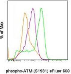
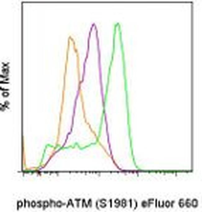
- Experimental details
- Normal human peripheral blood cells untreated (orange histogram), treated with Anti-Human CD3 and CD28 Functional Grade Purifieds (Product # 16-0037-81 and Product # 16-0289-81) (purple histogram), or treated with Anti-Human CD3 and CD28 Functional Grade Purifieds and Etopside (green histogram) were stained intracellularly with Anti-Human/Mouse phospho-ATM (S1981) eFluor® 660 using the Intracellular Fixation & Permeabilization Buffer Set (Product # 88-8824-00) and protocol. Cells in the lymphocyte gate were used for analysis.