Explore
 Validate
 Learn
Learn Western blot
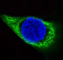
 Immunocytochemistry
Antibody data
- Antibody Data
- Antigen structure
- References [1]
- Comments [0]
- Validations
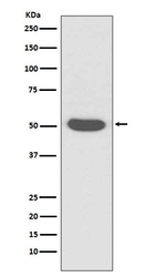
- Western blot [1]
Submit
Validation data
Reference
Comment
Report error
- Product number
- M01432-2 - Provider product page

- Provider
- Boster Biological Technology
- Product name
- Anti-Cytokeratin 14 KRT14 Rabbit Monoclonal Antibody
- Antibody type
- Monoclonal
- Description
- Monoclonal antibody for Cytokeratin 14/KRT14 detection. Host: Rabbit.Size: 100ug/vial. Tested applications: IF, IHC, ICC, WB. Reactive species: Human, Mouse, Rat Cytokeratin 14/KRT14 information: Molecular Weight: 51561 MW; Subcellular Localization: Cytoplasm. Nucleus. Expressed in both as a filamentous pattern; Tissue Specificity: Detected in the basal layer, lowered within the more apically located layers specifically in the stratum spinosum, stratum granulosum but is not detected in stratum corneum. Strongly expressed in the outer root sheath of anagen follicles but not in the germinative matrix, inner root sheath or hair. Found in keratinocytes surrounding the club hair during telogen.
- Reactivity
- Human, Mouse, Rat
- Host
- Rabbit
- Antibody clone number
- FEF-11
- Vial size
- 100ug/vial
- Concentration
- 0.5-1mg/ml, actual concentration vary by lot. Use suggested dilution ratio to decide dilution procedure.
- Storage
- At -20°C for one year. Avoid repeated freezing and thawing.
Submitted references Improvement of a Three-Layered in vitro Skin Model for Topical Application of Irritating Substances.
Schmidt FF, Nowakowski S, Kluger PJ
Frontiers in bioengineering and biotechnology 2020;8:388
Frontiers in bioengineering and biotechnology 2020;8:388
No comments: Submit comment
Supportive validation
- Submitted by
- Boster Biological Technology (provider)
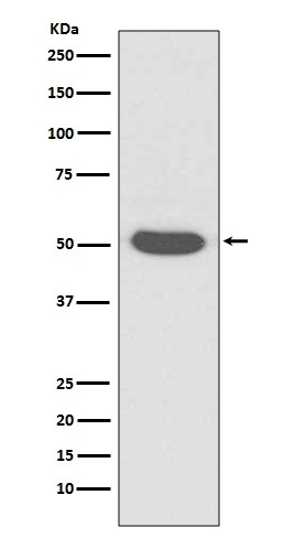
- Main image
- Experimental details
- Western blot analysis of Cytokeratin 14 expression in A431 cell lysate (M01432-2). Electrophoresis was performed on a 5-20% SDS-PAGE gel at 70V (Stacking gel) / 90V (Resolving gel) for 2-3 hours. The sample well of each lane was loaded with 50ug of sample under reducing conditions. After Electrophoresis, proteins were transferred to a Nitrocellulose membrane at 150mA for 50-90 minutes. Blocked the membrane with 5% Non-fat Milk/ TBS for 1.5 hour at RT. The membrane was incubated with rabbit anti-KRT14 monoclonal antibody (Catalog # M01432-2) overnight at 4°C, then washed with TBS-0.1%Tween 3 times with 5 minutes each and probed with a goat anti-rabbit IgG-HRP secondary antibody at a dilution of 1:10000 for 1.5 hour at RT. The signal is developed using an Enhanced Chemiluminescent detection (ECL) kit (Catalog # EK1002) with Tanon 5200 system. A specific band was detected for KRT14
- Additional image