 Explore
 Validate
 Learn
Learn Western blot
 Immunohistochemistry
Antibody data
- Antibody Data
- Antigen structure
- References [23]
- Comments [0]
- Validations
- Western blot [2]
- Flow cytometry [1]
- Other assay [5]
Submit
Validation data
Reference
Comment
Report error
- Product number
- PA5-16722 - Provider product page

- Provider
- Invitrogen Antibodies
- Product name
- Cytokeratin 14 Polyclonal Antibody
- Antibody type
- Polyclonal
- Antigen
- Synthetic peptide
- Description
- PA5-16722 targets Cytokeratin 14 in immunohistochemistry (paraffin and Western blot applications and shows reactivity with Human samples. The PA5-16722 immunogen is a synthetic peptide derived from C-terminal of human Keratin 14.
- Reactivity
- Human, Mouse
- Host
- Rabbit
- Isotype
- IgG
- Vial size
- 500 µL
- Concentration
- 0.235 mg/mL
- Storage
- -20° C, Avoid Freeze/Thaw Cycles
Submitted references SARS-CoV-2 Infection Dysregulates Cilia and Basal Cell Homeostasis in the Respiratory Epithelium of Hamsters.
EZH2 and Endometrial Cancer Development: Insights from a Mouse Model.
Functional similarity between TGF-beta type 2 and type 1 receptors in the female reproductive tract.
Stress keratin 17 enhances papillomavirus infection-induced disease by downregulating T cell recruitment.
Enhancer of Zeste 2 Polycomb Repressive Complex 2 Subunit Is Required for Uterine Epithelial Integrity.
Microenvironment-Induced Non-sporadic Expression of the AXL and cKIT Receptors Are Related to Epithelial Plasticity and Drug Resistance.
Hyaluronan Rich Microenvironment in the Limbal Stem Cell Niche Regulates Limbal Stem Cell Differentiation.
The Hippo kinases LATS1 and 2 control human breast cell fate via crosstalk with ERα.
Constitutively activated PI3K accelerates tumor initiation and modifies histopathology of breast cancer.
PIK3CA(H1047R) induces multipotency and multi-lineage mammary tumours.
Programmed synthesis of three-dimensional tissues.
Aspirin blocks growth of breast tumor cells and tumor-initiating cells and induces reprogramming factors of mesenchymal to epithelial transition.
Modelling breast cancer requires identification and correction of a critical cell lineage-dependent transduction bias.
Afadin requirement for cytokine expressions in keratinocytes during chemically induced inflammation in mice.
Label-retaining, quiescent globose basal cells are found in the olfactory epithelium.
Solitary tumours associated with Jaagsiekte retrovirus in sheep are heterogeneous and contain cells expressing markers identifying progenitor cells in lung repair.
A small molecule inhibitor of SRC family kinases promotes simple epithelial differentiation of human pluripotent stem cells.
Global expression profiling of globose basal cells and neurogenic progression within the olfactory epithelium.
Ascl1 (Mash1) knockout perturbs differentiation of nonneuronal cells in olfactory epithelium.
Py2T murine breast cancer cells, a versatile model of TGFβ-induced EMT in vitro and in vivo.
An immunohistochemical method to study breast cancer cell subpopulations and their growth regulation by hormones in three-dimensional cultures.
Luminal expression of PIK3CA mutant H1047R in the mammary gland induces heterogeneous tumors.
An oestrogen-dependent model of breast cancer created by transformation of normal human mammary epithelial cells.
Schreiner T, Allnoch L, Beythien G, Marek K, Becker K, Schaudien D, Stanelle-Bertram S, Schaumburg B, Mounogou Kouassi N, Beck S, Zickler M, Gabriel G, Baumgärtner W, Armando F, Ciurkiewicz M
International journal of molecular sciences 2022 May 4;23(9)
International journal of molecular sciences 2022 May 4;23(9)
EZH2 and Endometrial Cancer Development: Insights from a Mouse Model.
Fang X, Ni N, Wang X, Tian Y, Ivanov I, Rijnkels M, Bayless KJ, Lydon JP, Li Q
Cells 2022 Mar 7;11(5)
Cells 2022 Mar 7;11(5)
Functional similarity between TGF-beta type 2 and type 1 receptors in the female reproductive tract.
Ni N, Fang X, Li Q
Scientific reports 2021 Apr 29;11(1):9294
Scientific reports 2021 Apr 29;11(1):9294
Stress keratin 17 enhances papillomavirus infection-induced disease by downregulating T cell recruitment.
Wang W, Uberoi A, Spurgeon M, Gronski E, Majerciak V, Lobanov A, Hayes M, Loke A, Zheng ZM, Lambert PF
PLoS pathogens 2020 Jan;16(1):e1008206
PLoS pathogens 2020 Jan;16(1):e1008206
Enhancer of Zeste 2 Polycomb Repressive Complex 2 Subunit Is Required for Uterine Epithelial Integrity.
Fang X, Ni N, Lydon JP, Ivanov I, Bayless KJ, Rijnkels M, Li Q
The American journal of pathology 2019 Jun;189(6):1212-1225
The American journal of pathology 2019 Jun;189(6):1212-1225
Microenvironment-Induced Non-sporadic Expression of the AXL and cKIT Receptors Are Related to Epithelial Plasticity and Drug Resistance.
Jokela TA, Engelsen AST, Rybicka A, Pelissier Vatter FA, Garbe JC, Miyano M, Tiron C, Ferariu D, Akslen LA, Stampfer MR, Lorens JB, LaBarge MA
Frontiers in cell and developmental biology 2018;6:41
Frontiers in cell and developmental biology 2018;6:41
Hyaluronan Rich Microenvironment in the Limbal Stem Cell Niche Regulates Limbal Stem Cell Differentiation.
Gesteira TF, Sun M, Coulson-Thomas YM, Yamaguchi Y, Yeh LK, Hascall V, Coulson-Thomas VJ
Investigative ophthalmology & visual science 2017 Sep 1;58(11):4407-4421
Investigative ophthalmology & visual science 2017 Sep 1;58(11):4407-4421
The Hippo kinases LATS1 and 2 control human breast cell fate via crosstalk with ERα.
Britschgi A, Duss S, Kim S, Couto JP, Brinkhaus H, Koren S, De Silva D, Mertz KD, Kaup D, Varga Z, Voshol H, Vissieres A, Leroy C, Roloff T, Stadler MB, Scheel CH, Miraglia LJ, Orth AP, Bonamy GM, Reddy VA, Bentires-Alj M
Nature 2017 Jan 26;541(7638):541-545
Nature 2017 Jan 26;541(7638):541-545
Constitutively activated PI3K accelerates tumor initiation and modifies histopathology of breast cancer.
Sheen MR, Marotti JD, Allegrezza MJ, Rutkowski M, Conejo-Garcia JR, Fiering S
Oncogenesis 2016 Oct 31;5(10):e267
Oncogenesis 2016 Oct 31;5(10):e267
PIK3CA(H1047R) induces multipotency and multi-lineage mammary tumours.
Koren S, Reavie L, Couto JP, De Silva D, Stadler MB, Roloff T, Britschgi A, Eichlisberger T, Kohler H, Aina O, Cardiff RD, Bentires-Alj M
Nature 2015 Sep 3;525(7567):114-8
Nature 2015 Sep 3;525(7567):114-8
Programmed synthesis of three-dimensional tissues.
Todhunter ME, Jee NY, Hughes AJ, Coyle MC, Cerchiari A, Farlow J, Garbe JC, LaBarge MA, Desai TA, Gartner ZJ
Nature methods 2015 Oct;12(10):975-81
Nature methods 2015 Oct;12(10):975-81
Aspirin blocks growth of breast tumor cells and tumor-initiating cells and induces reprogramming factors of mesenchymal to epithelial transition.
Maity G, De A, Das A, Banerjee S, Sarkar S, Banerjee SK
Laboratory investigation; a journal of technical methods and pathology 2015 Jul;95(7):702-17
Laboratory investigation; a journal of technical methods and pathology 2015 Jul;95(7):702-17
Modelling breast cancer requires identification and correction of a critical cell lineage-dependent transduction bias.
Hines WC, Yaswen P, Bissell MJ
Nature communications 2015 Apr 21;6:6927
Nature communications 2015 Apr 21;6:6927
Afadin requirement for cytokine expressions in keratinocytes during chemically induced inflammation in mice.
Yoshida T, Iwata T, Takai Y, Birchmeier W, Yamato M, Okano T
Genes to cells : devoted to molecular & cellular mechanisms 2014 Nov;19(11):842-52
Genes to cells : devoted to molecular & cellular mechanisms 2014 Nov;19(11):842-52
Label-retaining, quiescent globose basal cells are found in the olfactory epithelium.
Jang W, Chen X, Flis D, Harris M, Schwob JE
The Journal of comparative neurology 2014 Mar;522(4):731-49
The Journal of comparative neurology 2014 Mar;522(4):731-49
Solitary tumours associated with Jaagsiekte retrovirus in sheep are heterogeneous and contain cells expressing markers identifying progenitor cells in lung repair.
De las Heras M, de Martino A, Borobia M, Ortín A, Álvarez R, Borderías L, Giménez-Más JA
Journal of comparative pathology 2014 Feb-Apr;150(2-3):138-47
Journal of comparative pathology 2014 Feb-Apr;150(2-3):138-47
A small molecule inhibitor of SRC family kinases promotes simple epithelial differentiation of human pluripotent stem cells.
Lian X, Selekman J, Bao X, Hsiao C, Zhu K, Palecek SP
PloS one 2013;8(3):e60016
PloS one 2013;8(3):e60016
Global expression profiling of globose basal cells and neurogenic progression within the olfactory epithelium.
Krolewski RC, Packard A, Schwob JE
The Journal of comparative neurology 2013 Mar 1;521(4):833-59
The Journal of comparative neurology 2013 Mar 1;521(4):833-59
Ascl1 (Mash1) knockout perturbs differentiation of nonneuronal cells in olfactory epithelium.
Krolewski RC, Packard A, Jang W, Wildner H, Schwob JE
PloS one 2012;7(12):e51737
PloS one 2012;7(12):e51737
Py2T murine breast cancer cells, a versatile model of TGFβ-induced EMT in vitro and in vivo.
Waldmeier L, Meyer-Schaller N, Diepenbruck M, Christofori G
PloS one 2012;7(11):e48651
PloS one 2012;7(11):e48651
An immunohistochemical method to study breast cancer cell subpopulations and their growth regulation by hormones in three-dimensional cultures.
Pinto MP, Jacobsen BM, Horwitz KB
Frontiers in endocrinology 2011;2:15
Frontiers in endocrinology 2011;2:15
Luminal expression of PIK3CA mutant H1047R in the mammary gland induces heterogeneous tumors.
Meyer DS, Brinkhaus H, Müller U, Müller M, Cardiff RD, Bentires-Alj M
Cancer research 2011 Jul 1;71(13):4344-51
Cancer research 2011 Jul 1;71(13):4344-51
An oestrogen-dependent model of breast cancer created by transformation of normal human mammary epithelial cells.
Duss S, André S, Nicoulaz AL, Fiche M, Bonnefoi H, Brisken C, Iggo RD
Breast cancer research : BCR 2007;9(3):R38
Breast cancer research : BCR 2007;9(3):R38
No comments: Submit comment
Supportive validation
- Submitted by
- Invitrogen Antibodies (provider)
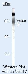
- Main image
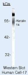
- Experimental details
- Western blot of Cytokeratin 14 using Cytokeratin 14 Polyclonal Antibody (Product # PA5-16722) on LS174T Cells.
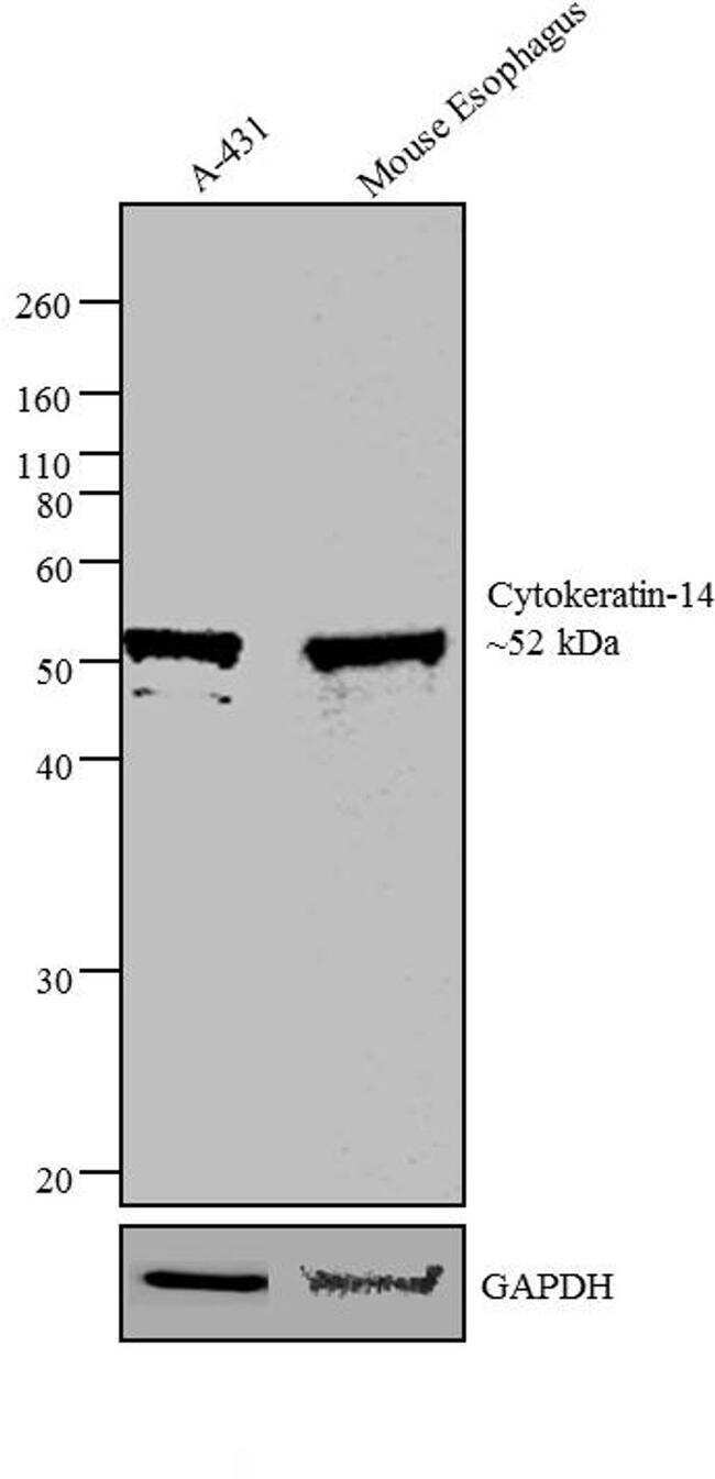
- Submitted by
- Invitrogen Antibodies (provider)
- Main image
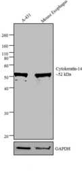
- Experimental details
- Western blot analysis was performed on whole cell extract and tissue extract of A-431 (Lane 1) and Mouse Esophagus Tissue (Lane 2). The blots were probed with Anti-Cytokeratin 14 Rabbit Polyclonal Antibody (Product # PA5-16722, 1 µg/mL) and detected by chemiluminescence using Goat anti-Rabbit IgG (H+L) Superclonal™ Secondary Antibody, HRP conjugate (Product # A27036, 0.4 µg/mL, 1:2500 dilution). A ~ 52 kDa band corresponding to Cytokeratin 14 was observed in the cell line and tissue tested. Known quantity of protein samples were electrophoresed using Novex® NuPAGE® 4-12 % Bis-Tris gel (Product # NP0321BOX), XCell SureLock™ Electrophoresis System (Product # EI0002) and Novex® Sharp Pre-Stained Protein Standard (Product # LC5800). Resolved proteins were then transferred onto a nitrocellulose membrane with iBlot® 2 Dry Blotting System (Product # IB21001). The membrane was probed with the relevant primary and secondary Antibody following blocking with 5 % skimmed milk. Chemiluminescent detection was performed using Pierce™ ECL Western Blotting Substrate (Product # 32106).
Supportive validation
- Submitted by
- Invitrogen Antibodies (provider)
- Main image
- Experimental details
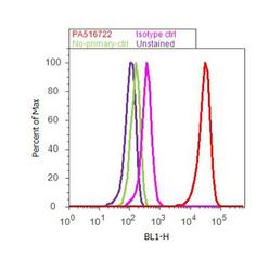
- Flow cytometry analysis of Cytokeratin 14 was done on A-431 cells. Cells were fixed with 70% ethanol for 10 minutes, permeabilized with 0.25% Triton™ X-100 for 20 minutes, and blocked with 5% BSA for 30 minutes at room temperature. Cells were labeled with Cytokeratin 14 Rabbit Polyclonal Antibody (PA5-16722, red histogram) or with rabbit isotype control (pink histogram) at 3-5 ug/million cells in 2.5% BSA. After incubation at room temperature for 2 hours, the cells were labeled with Alexa Fluor® 488 Goat Anti-Rabbit Secondary Antibody (A11008) at a dilution of 1:400 for 30 minutes at room temperature. The representative 10, 000 cells were acquired and analyzed for each sample using an Attune® Acoustic Focusing Cytometer. The purple histogram represents unstained control cells and the green histogram represents no-primary-antibody control.
Supportive validation
- Submitted by
- Invitrogen Antibodies (provider)
- Main image
- Experimental details
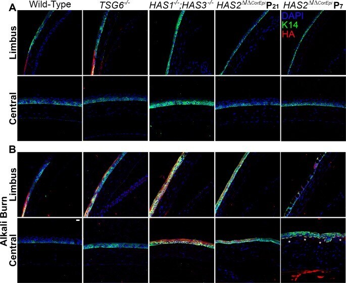
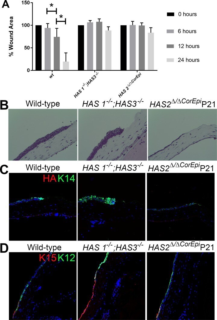
- Figure 7 Expression profiles of HA and K14 in the corneas of HAS1 -/- ;HAS3 -/- , HAS2 Delta/DeltaCorEpi , TSG-6 -/- , and wild-type mice. HA (red) and K14 (green) were stained in uninjured (A) and alkali burnt (B) corneas of HAS1 -/- ;HAS3 -/- , HAS2 Delta/DeltaCorEpi (induced at P21 and P7), TSG-6 -/- , and wild-type mice and images captured of the limbal region (Limbus) and central cornea (Central). The TSG-6 -/- and wild-type mice show HA staining in the corneal limbus, whereas HAS1 -/- ;HAS3 -/- and HAS2 Delta/DeltaCorEpi (induced at P21 and P7) mice lack HA staining in the limbus. Two weeks after alkali burn, TSG-6 -/- mice present an increase in HA expression into the peripheral cornea, HAS1 -/- ;HAS3 -/- and HAS2 Delta/DeltaCorEpi (induced at P21 and P7) mice express HA throughout the entire cornea, whereas wild-type mice present no changes in HA expression after injury. The dashed line (lower right image) shows the division between epithelial cells and the stroma and the asterisks mark where epithelial cells are growing into the stroma. Scale bar: 20 mum.
- Submitted by
- Invitrogen Antibodies (provider)
- Main image
- Experimental details
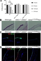
- Figure 9 HAS1 -/- ;HAS3 -/- and HAS2 Delta/DeltaCorEpi mice show delayed wound healing using an ex vivo debridement wound model. HAS1 -/- ;HAS3 -/- , HAS2 Delta/DeltaCorEpi (induced at P21), and wild-type mice were euthanized, debridement wounds were made on the central cornea, and eyeballs were removed and placed in culture. The rate of wound healing was assessed at 6, 12, and 24 hours by placing a drop of fluorescein on the eyeball and washing and capturing images using a fluorescence stereomicroscope. The wound area was measured and plotted in a graph as the percentage of wound area remaining (A). *P
- Submitted by
- Invitrogen Antibodies (provider)
- Main image
- Experimental details
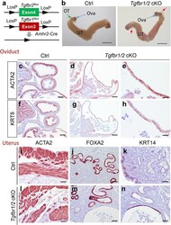
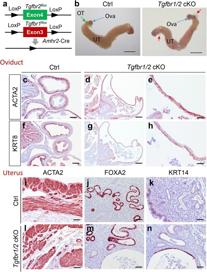
- Figure 6 Phenotypic characterization of Tgfbr1/2 cKO. ( a ) Schematic representation of generation of Tgfbr1/2 cKO. ( b ) Gross image of uteri from controls and Tgfbr1/2 cKO at 3 months of age. UT, uterus; OT, oviduct; Ova, ovary. Scale bar = 4 mm. ( c-h ) Localization of ACTA2 and KRT8 in the oviducts of controls and Tgfbr1/2 cKO at 3 months of age. ( e , h ) Are higher magnification images for ( d , g ). ( i-n ) Immunostaining of ACTA2, FOXA2, and KRT14 using uteri from controls and Tgfbr1/2 cKO at 3 months of age. Three independent mice were examined for each genotype. Scale bar equals 25 um ( e , h ), 50 um ( i - n ), 100 um ( c , d , f , g ).
- Submitted by
- Invitrogen Antibodies (provider)
- Main image

- Experimental details
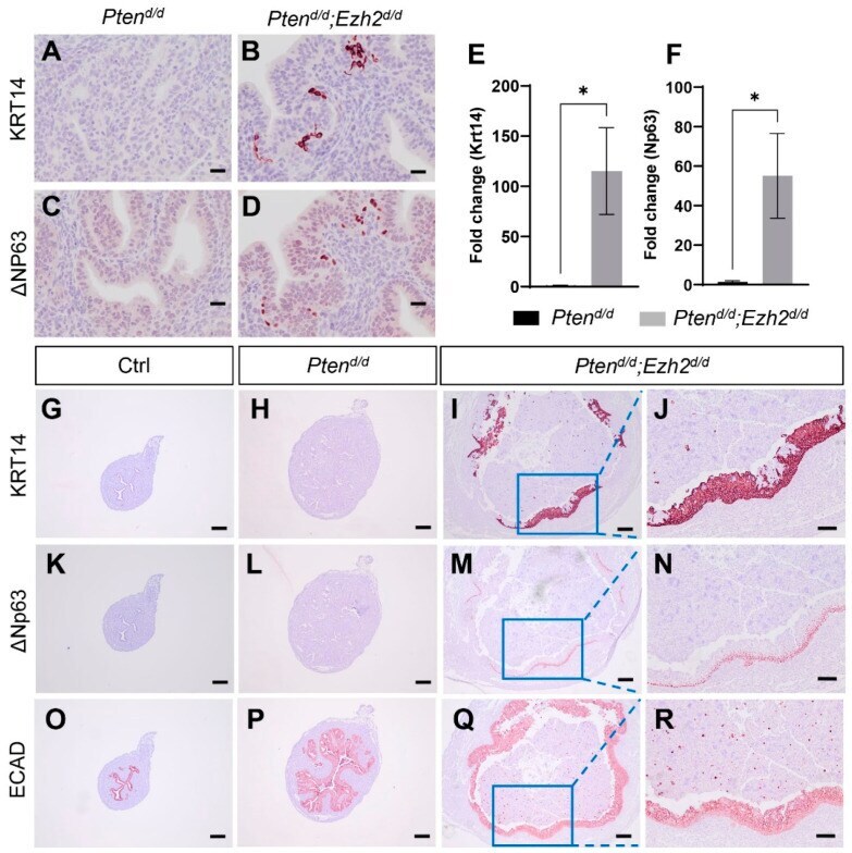
- Deletion of Ezh2 intensifies epithelial stratification in PTEN-depleted uteri. ( A - D ) Immunostaining of KRT14 and DeltaNP63 using uteri from three-week-old Pten d/d and Pten d/d ; Ezh2 d/d mice. ( E , F ) Transcript levels of Krt14 and Delta Np63 in uterine epithelia isolated from three-week-old Pten d/d and Pten d/d ; Ezh2 d/d mice. n = 4-5. Data are mean +- s.e.m. * p < 0.05. Note that one sample from the Pten d/d group had undetectable Delta Np63 expression and was not included in panel ( F ). ( G - R ) Immunostaining of KRT14 ( G - J ), DeltaNP63 ( K - N ), and ECAD ( O - R ) in uteri from one-month-old Pten f/f (Ctrl), Pten d/d , and Pten d/d ; Ezh2 d/d mice. Panels ( J , N , R ) represent high power images of the boxed areas of panels ( I , M , Q ), respectively. At least three independent samples were examined for each genotype. Scale bar = 20 um ( A - D ), 100 um ( J , N , R ), and 200 um ( G - I , K - M , O - Q ).
- Submitted by
- Invitrogen Antibodies (provider)
- Main image
- Experimental details
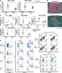
- Fig 4 Lack of stress keratin 17 leads to increased infiltration of CD8 + T cells and increased expression of IFNg-related genes. Mouse ears were infected with 10 9 VGE MmuPV1(Prep#1) per site. A) Percentage of CD8 + and CD4 + T cells based on flow cytometry analysis of normal ears (circles) versus ear MmuPv1 papillomas (squares) 4 weeks post-infection. For quantification of other populations, see S4A Fig . B) Immunofluorescent staining of frozen tissue sections of ear papillomas at 4 weeks post-infection. Green: CD8; Red: keratin 14 (K14); Blue: DAPI. Representative image for n = 4 samples (K17KO) and n = 6 samples (WT). C) RT-qPCR analysis of bulk RNA in mock-infected ears (circles) and ear MmuPV1 papillomas (squares) from K17KO and WT tissue 4 weeks post-infection. K17 expression was measured by TaqMan probe. IFNgamma, PDL1, CXCL9 and CXCL10 were measured by SYBR Green method. GAPDH was used for normalization. Data are represented as mean +- SEM. 2-way ANOVA Sidak multiple comparison was used. **p
