Explore
 Validate
 Learn
Learn Western blot
Antibody data
- Antibody Data
- Antigen structure
- References [3]
- Comments [0]
- Validations
- Western blot [3]
- Immunocytochemistry [1]
- Immunohistochemistry [1]
- Other assay [1]
Submit
Validation data
Reference
Comment
Report error
- Product number
- PA5-28002 - Provider product page

- Provider
- Invitrogen Antibodies
- Product name
- Cytokeratin 14 Polyclonal Antibody
- Antibody type
- Polyclonal
- Antigen
- Recombinant protein fragment
- Description
- Recommended positive controls: HeLa, rat olfactory epithelium. Predicted reactivity: Mouse (95%), Rat (94%), Dog (96%), Bovine (96%). Store product as a concentrated solution. Centrifuge briefly prior to opening the vial.
- Reactivity
- Human, Rat
- Host
- Rabbit
- Isotype
- IgG
- Vial size
- 100 µL
- Concentration
- 0.48 mg/mL
- Storage
- Store at 4°C short term. For long term storage, store at -20°C, avoiding freeze/thaw cycles.
Submitted references The microRNA/TET3/REST axis is required for olfactory globose basal cell proliferation and male behavior.
High-resolution 3D imaging of fixed and cleared organoids.
Human polyomavirus 6 and 7 are associated with pruritic and dyskeratotic dermatoses.
Yang D, Wu X, Zhou Y, Wang W, Wang Z
EMBO reports 2020 Sep 3;21(9):e49431
EMBO reports 2020 Sep 3;21(9):e49431
High-resolution 3D imaging of fixed and cleared organoids.
Dekkers JF, Alieva M, Wellens LM, Ariese HCR, Jamieson PR, Vonk AM, Amatngalim GD, Hu H, Oost KC, Snippert HJG, Beekman JM, Wehrens EJ, Visvader JE, Clevers H, Rios AC
Nature protocols 2019 Jun;14(6):1756-1771
Nature protocols 2019 Jun;14(6):1756-1771
Human polyomavirus 6 and 7 are associated with pruritic and dyskeratotic dermatoses.
Nguyen KD, Lee EE, Yue Y, Stork J, Pock L, North JP, Vandergriff T, Cockerell C, Hosler GA, Pastrana DV, Buck CB, Wang RC
Journal of the American Academy of Dermatology 2017 May;76(5):932-940.e3
Journal of the American Academy of Dermatology 2017 May;76(5):932-940.e3
No comments: Submit comment
Supportive validation
- Submitted by
- Invitrogen Antibodies (provider)
- Main image
- Experimental details
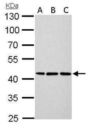
- Western blot analysis of Cytokeratin 14 using A) 30 µg Neuro2A whole cell lysate (B) 30 µg GL261 whole cell lysate and C) 30 µg C8D30 whole cell lysate. Samples were loaded onto a 10% SDS-PAGE gel and probed with a Cytokeratin 14 polyclonal antibody (Product # PA5-28002) at a dilution of 1:500.
- Submitted by
- Invitrogen Antibodies (provider)
- Main image

- Experimental details
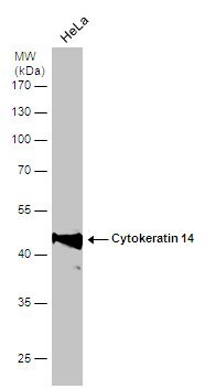
- Western blot analysis of Cytokeratin 14 using 30 µg of HeLa S3 lysate. Samples were loaded onto a 7.5% SDS-PAGE gel and probed with a Cytokeratin 14 polyclonal antibody (Product # PA5-28002) at a dilution of 1:3000.

- Submitted by
- Invitrogen Antibodies (provider)
- Main image

- Experimental details
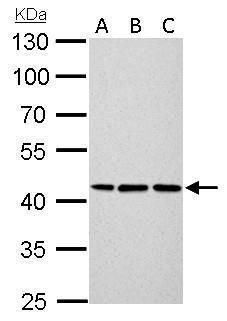
- Western Blot analysis of Cytokeratin 14 was performed by separating 30 µg of whole cell extract by 10% SDS-PAGE. Proteins were transferred to a membrane and probed with a Cytokeratin 14 Polyclonal Antibody (Product # PA5-28002) at a dilution of 1:6000.
Supportive validation
- Submitted by
- Invitrogen Antibodies (provider)
- Main image
- Experimental details
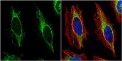
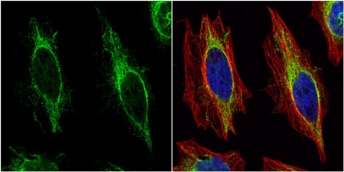
- Immunocytochemistry-Immunofluorescence analysis of Cytokeratin 14 was performed in HeLa cells fixed in 4% paraformaldehyde at RT for 15 min. Green: Cytokeratin 14 Polyclonal Antibody (Product # PA5 28002) diluted at 1:200. Red: alpha Tubulin, a cytoskeleton marker. Blue: Hoechst 33342 staining.
Supportive validation
- Submitted by
- Invitrogen Antibodies (provider)
- Main image
- Experimental details
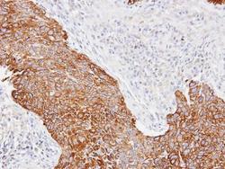
- Immunohistochemical analysis of paraffin-embedded Lung CA, using Cytokeratin 14 (Product # PA5-28002) antibody at 1:100 dilution. Antigen Retrieval: EDTA based buffer, pH 8.0, 15 min.

Supportive validation
- Submitted by
- Invitrogen Antibodies (provider)
- Main image
- Experimental details
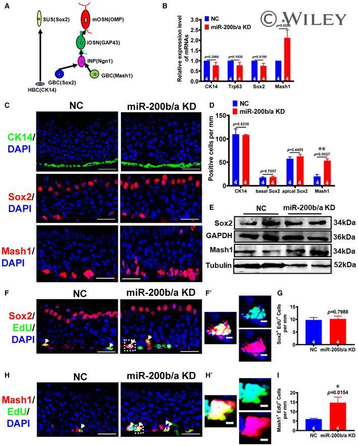
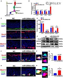
- miR-200b/a are crucial for the proliferation of the Mash1-marked GBC s Schematic illustration of developmentally expressed olfactory cell types: horizontal basal cell (HBC), globose basal cell (GBC), intermediate nerve progenitor (INP), and immature olfactory sensory neuron (iOSN), mature olfactory sensory neuron (mOSN), and sustentacular cell (Sus). The mRNA levels of CK14, Trp63, Sox2, and Mash1 in the MOE of the NC and miR-200b/a KD mice, as revealed by qPCR analysis ( n = 4 mice each group; data represent the mean +- SEM; CK14: P = 0.2135, Trp63: P = 0.1930, Sox2: P = 0.5190, * P < 0.05; Student's t -test). Representative IF staining for CK14, Sox2, and Mash1 in the MOE of the NC and miR-200b/a KD mice. Scale bars, 20 mum. Quantification of the number of CK14 + , basal Sox2 + , apical Sox2 + , and Mash1 + cells in the MOE of the NC and miR-200b/a KD mice ( n = 4 mice each group; data represent the mean +- SEM; CK14: P = 0.8998, basal Sox2: P = 0.7056, apical Sox2: P = 0.4455, ** P < 0.01; Student's t -test). Western blot analysis of the Sox2 and Mash1 protein levels in the MOE of the NC and miR-200b/a KD mice ( n = 2 mice each group). GAPDH and Tubulin served as loading controls. The molecular weight of each band is indicated at the right. Representative IF costaining with Sox2 antibody and EdU in the MOE of the NC and miR-200b/a KD mice. The white arrows indicate the cells positive for both Sox2 and EdU. (F') is shown at higher magnification of the boxed region, as three