 Explore
 Validate
 Learn
Learn Western blot
 Immunocytochemistry
Antibody data
- Antibody Data
- Antigen structure
- References [1]
- Comments [0]
- Validations
- Immunocytochemistry [1]
Submit
Validation data
Reference
Comment
Report error
- Product number
- MAB3164 - Provider product page

- Provider
- R&D Systems
- Product name
- Human Cytokeratin 14 Antibody
- Antibody type
- Monoclonal
- Description
- Protein A or G purified from hybridoma culture supernatant. Detects human Cytokeratin 14 in Western blots.
- Reactivity
- Human
- Host
- Mouse
- Conjugate
- Unconjugated
- Isotype
- IgG
- Antibody clone number
- LL001
- Vial size
- 100 ug
- Concentration
- LYOPH
- Storage
- Use a manual defrost freezer and avoid repeated freeze-thaw cycles. 12 months from date of receipt, -20 to -70 °C as supplied. 1 month, 2 to 8 °C under sterile conditions after reconstitution. 6 months, -20 to -70 °C under sterile conditions after reconstitution.
Submitted references Decreased IGF type 1 receptor signaling in mammary epithelium during pregnancy leads to reduced proliferation, alveolar differentiation, and expression of insulin receptor substrate (IRS)-1 and IRS-2.
Sun Z, Shushanov S, LeRoith D, Wood TL
Endocrinology 2011 Aug;152(8):3233-45
Endocrinology 2011 Aug;152(8):3233-45
No comments: Submit comment
Supportive validation
- Submitted by
- R&D Systems (provider)
- Main image
- Experimental details
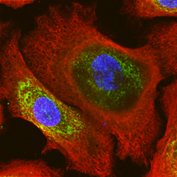
- Cytokeratin 14 in NHEK Human Normal Epidermal Keratinocytes. Cytokeratin 14 was detected in immersion fixed NHEK human normal epidermal keratinocytes using Mouse Anti-Human Cytokeratin 14 Monoclonal Antibody (Catalog # MAB3164) at 10 µg/mL for 3 hours at room temperature. Cells were stained using the NorthernLights™ 557-conjugated Anti-Mouse IgG Secondary Antibody (red; Catalog # NL007). CKAP4/p63 was also detected using Sheep Anti-Human CKAP4/p63 Affinity-purified Polyclonal Antibody (Catalog # AF7355) and stained using the NorthernLights™ 493-conjugated Anti-Sheep IgG Secondary Antibody (green; Catalog # NL012). Cells were counterstained with DAPI (blue). Specific staining of Cytokeratin 14 was localized to intermediate filaments. View our protocol for Fluorescent ICC Staining of Cells on Coverslips.
