 Explore
 Validate
 Learn
Learn Western blot
 Immunocytochemistry
Antibody data
- Antibody Data
- Antigen structure
- References [1]
- Comments [0]
- Validations
- Immunocytochemistry [1]
- Immunohistochemistry [1]
Submit
Validation data
Reference
Comment
Report error
- Product number
- HPA024554 - Provider product page

- Provider
- Atlas Antibodies
- Proper citation
- Atlas Antibodies Cat#HPA024554, RRID:AB_1852207
- Product name
- Anti-KRT15
- Antibody type
- Polyclonal
- Description
- Polyclonal Antibody against Human KRT15, Gene description: keratin 15, Alternative Gene Names: CK15, K15, K1CO, Validated applications: WB, IHC, ICC, Uniprot ID: P19012, Storage: Store at +4°C for short term storage. Long time storage is recommended at -20°C.
- Reactivity
- Human
- Host
- Rabbit
- Conjugate
- Unconjugated
- Isotype
- IgG
- Vial size
- 100 µl
- Concentration
- 0.1 mg/ml
- Storage
- Store at +4°C for short term storage. Long time storage is recommended at -20°C.
- Handling
- The antibody solution should be gently mixed before use.
Submitted references The value of desmosomal plaque-related markers to distinguish squamous cell carcinoma and adenocarcinoma of the lung
Galindo I, Gómez-Morales M, Díaz-Cano I, Andrades Á, Caba-Molina M, Miranda-León M, Medina P, Martín-Padron J, Fárez-Vidal M
Upsala Journal of Medical Sciences 2019;125(1):19-29
Upsala Journal of Medical Sciences 2019;125(1):19-29
No comments: Submit comment
Supportive validation
- Submitted by
- Atlas Antibodies (provider)
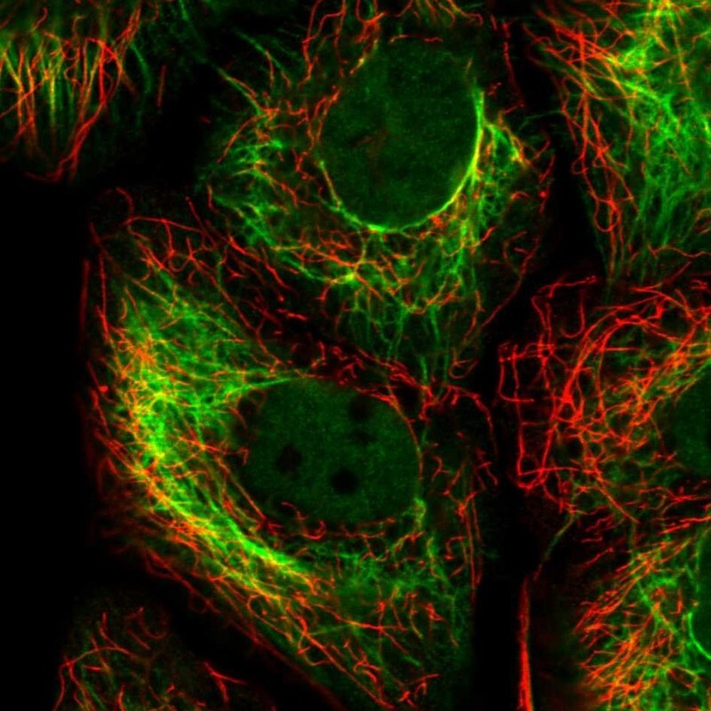
- Main image

- Experimental details
- Immunofluorescent staining of human cell line HaCaT shows localization to nucleoplasm & intermediate filaments.
- Sample type
- Human
Supportive validation
- Submitted by
- Atlas Antibodies (provider)
- Enhanced method
- Orthogonal validation
- Main image
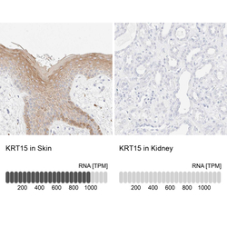
- Experimental details
- Immunohistochemistry analysis in human skin and kidney tissues using HPA024554 antibody. Corresponding KRT15 RNA-seq data are presented for the same tissues.
- Sample type
- Human
- Protocol
- Protocol
