 Explore
 Validate
 Learn
Learn Western blot
Antibody data
- Antibody Data
- Antigen structure
- References [17]
- Comments [0]
- Validations
- Western blot [2]
- Immunohistochemistry [1]
- Flow cytometry [1]
- Other assay [2]
Submit
Validation data
Reference
Comment
Report error
- Product number
- MA5-13730 - Provider product page

- Provider
- Invitrogen Antibodies
- Product name
- Cytokeratin 16 Monoclonal Antibody (LL025)
- Antibody type
- Monoclonal
- Antigen
- Synthetic peptide
- Description
- MA5-13730 targets Cytokeratin 16 in immunofluorescence and immunohistochemistry (paraffin) applications and shows reactivity with Human samples. Staining of formalin/paraffin tissues requires boiling tissue sections in 10 mM citrate buffer, pH 6.0 for 10-20 minutes followed by cooling at room temperature for 20 minutes.
- Antibody clone number
- LL025
- Concentration
- 0.2 mg/mL
Submitted references Expression of keratins 8, 18, and 19 in epithelia of atrophic oral lichen planus.
Epithelial Fli1 deficiency drives systemic autoimmunity and fibrosis: Possible roles in scleroderma.
The single-chain anti-TNF-α antibody DLX105 induces clinical and biomarker responses upon local administration in patients with chronic plaque-type psoriasis.
Development of full-thickness human skin equivalents with blood and lymph-like capillary networks by cell coating technology.
Interactions between myofibroblast differentiation and epidermogenesis in constructing human living skin equivalents.
Development of skin-humanized mouse models of pachyonychia congenita.
Down-regulation of keratin 4 and keratin 13 expression in oral squamous cell carcinoma and epithelial dysplasia: a clue for histopathogenesis.
Methods to promote Notch signaling at the biomaterial interface and evaluation in a rafted organ culture model.
Effects of antikeratin 16 antibodies on the expression of Toll-like receptors 2 and 4 in keratinocytes.
Keratinocyte migration, proliferation, and differentiation in chronic ulcers from patients with diabetes and normal wounds.
Proteomic analysis of scleroderma lesional skin reveals activated wound healing phenotype of epidermal cell layer.
Innate immune modulation of keratinocytes by antikeratin 16 antibodies.
Morphological evidence for the role of suprabasal keratinocytes in wound reepithelialization.
Mek1 alters epidermal growth and differentiation.
A study of cytokeratin profiles in localized cutaneous amyloids.
Organotypic keratinocyte coculture using normal human serum: an immunomorphological study at light and electron microscopic levels.
Organotypic keratinocyte coculture using normal human serum: an immunomorphological study at light and electron microscopic levels.
Schreurs O, Karatsaidis A, Balta MG, Grung B, Hals EKB, Schenck K
European journal of oral sciences 2020 Feb;128(1):7-17
European journal of oral sciences 2020 Feb;128(1):7-17
Epithelial Fli1 deficiency drives systemic autoimmunity and fibrosis: Possible roles in scleroderma.
Takahashi T, Asano Y, Sugawara K, Yamashita T, Nakamura K, Saigusa R, Ichimura Y, Toyama T, Taniguchi T, Akamata K, Noda S, Yoshizaki A, Tsuruta D, Trojanowska M, Sato S
The Journal of experimental medicine 2017 Apr 3;214(4):1129-1151
The Journal of experimental medicine 2017 Apr 3;214(4):1129-1151
The single-chain anti-TNF-α antibody DLX105 induces clinical and biomarker responses upon local administration in patients with chronic plaque-type psoriasis.
Tsianakas A, Brunner PM, Ghoreschi K, Berger C, Loser K, Röcken M, Stingl G, Luger T, Jung T
Experimental dermatology 2016 Jun;25(6):428-33
Experimental dermatology 2016 Jun;25(6):428-33
Development of full-thickness human skin equivalents with blood and lymph-like capillary networks by cell coating technology.
Matsusaki M, Fujimoto K, Shirakata Y, Hirakawa S, Hashimoto K, Akashi M
Journal of biomedical materials research. Part A 2015 Oct;103(10):3386-96
Journal of biomedical materials research. Part A 2015 Oct;103(10):3386-96
Interactions between myofibroblast differentiation and epidermogenesis in constructing human living skin equivalents.
Yang L, Hashimoto K, Tohyama M, Okazaki H, Dai X, Hanakawa Y, Sayama K, Shirakata Y
Journal of dermatological science 2012 Jan;65(1):50-7
Journal of dermatological science 2012 Jan;65(1):50-7
Development of skin-humanized mouse models of pachyonychia congenita.
García M, Larcher F, Hickerson RP, Baselga E, Leachman SA, Kaspar RL, Del Rio M
The Journal of investigative dermatology 2011 May;131(5):1053-60
The Journal of investigative dermatology 2011 May;131(5):1053-60
Down-regulation of keratin 4 and keratin 13 expression in oral squamous cell carcinoma and epithelial dysplasia: a clue for histopathogenesis.
Sakamoto K, Aragaki T, Morita K, Kawachi H, Kayamori K, Nakanishi S, Omura K, Miki Y, Okada N, Katsube K, Takizawa T, Yamaguchi A
Histopathology 2011 Mar;58(4):531-42
Histopathology 2011 Mar;58(4):531-42
Methods to promote Notch signaling at the biomaterial interface and evaluation in a rafted organ culture model.
Beckstead BL, Tung JC, Liang KJ, Tavakkol Z, Usui ML, Olerud JE, Giachelli CM
Journal of biomedical materials research. Part A 2009 Nov;91(2):436-46
Journal of biomedical materials research. Part A 2009 Nov;91(2):436-46
Effects of antikeratin 16 antibodies on the expression of Toll-like receptors 2 and 4 in keratinocytes.
Wu C, Luan Q, Li C, Zheng Z
Clinical and experimental dermatology 2009 Mar;34(2):236-9
Clinical and experimental dermatology 2009 Mar;34(2):236-9
Keratinocyte migration, proliferation, and differentiation in chronic ulcers from patients with diabetes and normal wounds.
Usui ML, Mansbridge JN, Carter WG, Fujita M, Olerud JE
The journal of histochemistry and cytochemistry : official journal of the Histochemistry Society 2008 Jul;56(7):687-96
The journal of histochemistry and cytochemistry : official journal of the Histochemistry Society 2008 Jul;56(7):687-96
Proteomic analysis of scleroderma lesional skin reveals activated wound healing phenotype of epidermal cell layer.
Aden N, Shiwen X, Aden D, Black C, Nuttall A, Denton CP, Leask A, Abraham D, Stratton R
Rheumatology (Oxford, England) 2008 Dec;47(12):1754-60
Rheumatology (Oxford, England) 2008 Dec;47(12):1754-60
Innate immune modulation of keratinocytes by antikeratin 16 antibodies.
Wu C, Li C, Wei L, Zheng Z
Experimental dermatology 2008 Aug;17(8):645-52
Experimental dermatology 2008 Aug;17(8):645-52
Morphological evidence for the role of suprabasal keratinocytes in wound reepithelialization.
Usui ML, Underwood RA, Mansbridge JN, Muffley LA, Carter WG, Olerud JE
Wound repair and regeneration : official publication of the Wound Healing Society [and] the European Tissue Repair Society 2005 Sep-Oct;13(5):468-79
Wound repair and regeneration : official publication of the Wound Healing Society [and] the European Tissue Repair Society 2005 Sep-Oct;13(5):468-79
Mek1 alters epidermal growth and differentiation.
Scholl FA, Dumesic PA, Khavari PA
Cancer research 2004 Sep 1;64(17):6035-40
Cancer research 2004 Sep 1;64(17):6035-40
A study of cytokeratin profiles in localized cutaneous amyloids.
Chang YT, Liu HN, Wang WJ, Lee DD, Tsai SF
Archives of dermatological research 2004 Jul;296(2):83-8
Archives of dermatological research 2004 Jul;296(2):83-8
Organotypic keratinocyte coculture using normal human serum: an immunomorphological study at light and electron microscopic levels.
Hinterhuber G, Marquardt Y, Diem E, Rappersberger K, Wolff K, Foedinger D
Experimental dermatology 2002 Oct;11(5):413-20
Experimental dermatology 2002 Oct;11(5):413-20
Organotypic keratinocyte coculture using normal human serum: an immunomorphological study at light and electron microscopic levels.
Hinterhuber G, Marquardt Y, Diem E, Rappersberger K, Wolff K, Foedinger D
Experimental dermatology 2002 Oct;11(5):413-20
Experimental dermatology 2002 Oct;11(5):413-20
No comments: Submit comment
Supportive validation
- Submitted by
- Invitrogen Antibodies (provider)
- Main image
- Experimental details
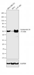
- Western blot analysis was performed on membrane enriched extracts (30 µg lysate) of MDA-MB-231 (Lane 1), HeLa (Lane 2) and U-87 MG (Lane 3). The blot was probed with Mouse Anti-Cytokeratin 16 Monoclonal Antibody (Product # MA5-13730, 2 µg/mL) and detected by chemiluminescence using Goat anti-Mouse IgG (H+L) Superclonal™ Secondary Antibody, HRP conjugate (Product # A28177, 0.4 µg/mL, 1:4000 dilution). A 51 kDa band corresponding to cytokeratin 16 was observed across the cell lines tested. Known quantity of protein samples were electrophoresed using Novex® NuPAGE® 4-12 % Bis-Tris gel (Product # NP0321BOX), XCell SureLock™ Electrophoresis System (Product # EI0002) and Novex® Sharp Pre-Stained Protein Standard (Product # LC5800). Resolved proteins were then transferred onto a nitrocellulose membrane with iBlot® 2 Dry Blotting System (Product # IB21001). The membrane was probed with the relevant primary and secondary Antibody using iBind™ Flex Western Starter Kit (Product # SLF2000S). Chemiluminescent detection was performed using Pierce™ ECL Western Blotting Substrate (Product # 32106).

- Submitted by
- Invitrogen Antibodies (provider)
- Main image

- Experimental details
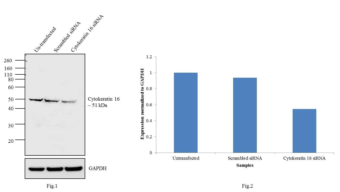
- Knockdown of Cytokeratin 16 was achieved by transfecting HeLa cells with Cytokeratin 16 specific validated siRNAs (Silencer® select Product # s7991). Western blot analysis (Fig. 1) was performed using whole cell extracts from the Cytokeratin 16 knockdown cells (lane 3), non-specific scrambled siRNA transfected cells (lane 2) and untransfected cells (lane 1). The blots were probed with Anti-Cytokeratin 16 Monoclonal Antibody (Product # MA5-13730, 1 µg/mL) and Goat anti-Mouse IgG (H+L) Superclonal™ Secondary Antibody, HRP conjugate (Product # A28177, 0.25 µg/mL, 1:4000 dilution). Densitometric analysis of this western blot is shown in histogram (Fig. 2). Decrease in signal upon siRNA mediated knock down confirms that antibody is specific to Cytokeratin 16.
Supportive validation
- Submitted by
- Invitrogen Antibodies (provider)
- Main image
- Experimental details
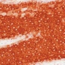
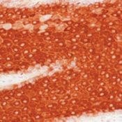
- Formalin-fixed, paraffin-embedded human squamous carcinoma of lung stained with Keratin-16 antibody using peroxidase-conjugate and AEC chromogen. Note cytoplasmic staining of tumor cells.
Supportive validation
- Submitted by
- Invitrogen Antibodies (provider)
- Main image

- Experimental details
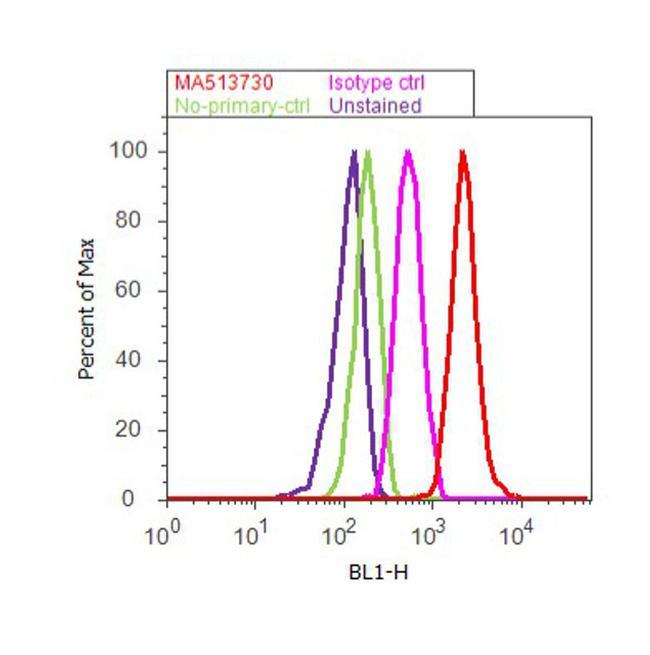
- Flow cytometry analysis of Cytokeratin 16 was done on A-431 cells. Cells were fixed with 70% ethanol for 10 minutes, permeabilized with 0.25% Triton™ X-100 for 20 minutes, and blocked with 5% BSA for 30 minutes at room temperature. Cells were labeled with Cytokeratin 16 Mouse Monoclonal Antibody (Product # MA5-13730, red histogram) or with mouse isotype control (pink histogram) at 3-5 µg/million cells in 2.5% BSA. After incubation at room temperature for 2 hours, the cells were labeled with Alexa Fluor® 488 Rabbit Anti-Mouse Secondary Antibody (Product # A11059) at a dilution of 1:400 for 30 minutes at room temperature. The representative 10, 000 cells were acquired and analyzed for each sample using an Attune® Acoustic Focusing Cytometer. The purple histogram represents unstained control cells and the green histogram represents no-primary-antibody control.
Supportive validation
- Submitted by
- Invitrogen Antibodies (provider)
- Main image
- Experimental details
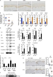
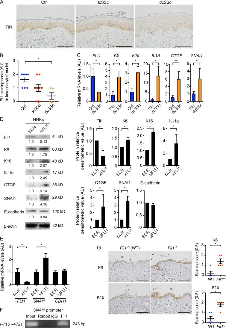
- Figure 1. Gene silencing of Fli1 leads to the induction of SSc-like protein and gene expression profiles in keratinocytes. (A) Immunohistochemistry for Fli1 on the skin sections of healthy controls ( n = 8), lcSSc patients with anticentromere antibody ( n = 8), and dcSSc patients with anti-topoisomerase I antibody ( n = 5). Representative images are shown. Bars, 100 um. (B) The results of Fli1 staining intensity in A were semiquantitatively evaluated with a four-point grading scale. Kruskal-Wallis test followed by Dunn's posthoc test was used. (C) mRNA was isolated from skin epidermal sheets from healthy controls ( n = 4) and dcSSc patients ( n = 6), and gene expressions were analyzed by qRT-PCR. (D) Whole cell lysates from cultured NHKs treated with control nonsilencing SCR or FLI1 siRNA (siFLI1) were subjected to immunoblotting. The values below each blot represent the relative levels of target molecules normalized by controls with densitometry. Representative images of the blots of four independent experiments are shown. (Right) The bar graphs summarize the relative values of the density from these four experiments. (E) The mRNA expression of FLI1 , SNAI1 , and CDH1 in NHKs treated with SCR or siFLI1 was evaluated. Relative expression levels from four independent experiments were normalized to the expression levels treated with SCR. (F) ChIP assay in NHKs was performed with anti-Fli1 antibody and the primers specific for the designated area of the SNAI1 gene promoter.
- Submitted by
- Invitrogen Antibodies (provider)
- Main image
- Experimental details
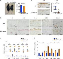
- Figure 2. K14Cre;fl/fl mice exhibit SSc-like epidermal phenotypes. (A, left) A representative image of mice at ~4 mo of age. (Right) The comparison of body weight between two strains of mice. n = 7 per genotype. (B, left) Immunohistochemistry for Fli1 in the back skin of mice. Bars, 50 um. (Right) The graph summarizes the result of semiquantitative scoring of the staining intensity. n = 7 per genotype. (C) Immunohistochemistry for K6, K16, IL-1alpha, CTGF, and SNAI1 in the back skin of mice. n = 5 per genotype. Bars, 50 um. (D) The results of staining intensity in the epidermis as shown in C were evaluated with semiquantitative scoring. n = 5 per genotype. (E) mRNA expressions of these key molecules were determined by qRT-PCR in the epidermal sheets prepared from the back skin of mice. n = 5 per genotype. Data are shown as mean +- SEM. *, P < 0.05; **, P < 0.01; by two-tailed Mann-Whitney U test. The results are from representative experiments that have been repeated twice in different pairs of mice with similar results. Representative images of immunohistochemistry are shown. AU, arbitrary units.
