Explore
 Validate
 Learn
LearnMA1-12294
antibody from Invitrogen Antibodies
Targeting: CDKN2B
CDK4I, INK4B, MTS2, P15, p15INK4b, TP15
 Western blot
 Immunocytochemistry
Antibody data
- Antibody Data
- Antigen structure
- References [3]
- Comments [0]
- Validations
- Immunocytochemistry [1]
- Other assay [4]
Submit
Validation data
Reference
Comment
Report error
- Product number
- MA1-12294 - Provider product page

- Provider
- Invitrogen Antibodies
- Product name
- CDKN2B Monoclonal Antibody (DCS114.1)
- Antibody type
- Monoclonal
- Antigen
- Recombinant full-length protein
- Reactivity
- Human
- Host
- Mouse
- Isotype
- IgG
- Antibody clone number
- DCS114.1
- Vial size
- 100 μg
- Concentration
- 1 mg/mL
- Storage
- -20°C, Avoid Freeze/Thaw Cycles
Submitted references DNMT1-Mediated DNA Methylation Targets CDKN2B to Promote the Repair of Retinal Ganglion Cells in Streptozotocin-Induced Mongolian Gerbils during Diabetic Retinopathy.
MiR-9-1 Suppresses Cell Proliferation and Promotes Apoptosis by Targeting UHRF1 in Lung Cancer.
Identification of Cyclobutane Pyrimidine Dimer-Responsive Genes Using UVB-Irradiated Human Keratinocytes Transfected with In Vitro-Synthesized Photolyase mRNA.
Wang X, Zhang J, Liao Y, Jin Y, Yu X, Li H, Yang Q, Li X, Chen R, Wu D, Zhu H
Computational and mathematical methods in medicine 2022;2022:9212116
Computational and mathematical methods in medicine 2022;2022:9212116
MiR-9-1 Suppresses Cell Proliferation and Promotes Apoptosis by Targeting UHRF1 in Lung Cancer.
Jia CY, Xiang W, Liu JB, Jiang GX, Sun F, Wu JJ, Yang XL, Xin R, Shi Y, Zhang DD, Li W, Zuberi Z, Zhang J, Lu GX, Wang HM, Wang PY, Yu F, Lv ZW, Ma YS, Fu D
Technology in cancer research & treatment 2021 Jan-Dec;20:15330338211041191
Technology in cancer research & treatment 2021 Jan-Dec;20:15330338211041191
Identification of Cyclobutane Pyrimidine Dimer-Responsive Genes Using UVB-Irradiated Human Keratinocytes Transfected with In Vitro-Synthesized Photolyase mRNA.
Boros G, Miko E, Muramatsu H, Weissman D, Emri E, van der Horst GT, Szegedi A, Horkay I, Emri G, Karikó K, Remenyik É
PloS one 2015;10(6):e0131141
PloS one 2015;10(6):e0131141
No comments: Submit comment
Supportive validation
- Submitted by
- Invitrogen Antibodies (provider)
- Main image
- Experimental details
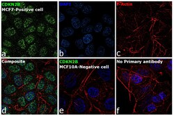
- Immunofluorescence analysis of CDKN2B was performed using 70% confluent log phase MCF7 cells. The cells were fixed with 4% paraformaldehyde for 10 minutes, permeabilized with 0.1% Triton™ X-100 for 15 minutes, and blocked with 2% BSA for 1 hour at room temperature. The cells were labeled with CDKN2B Mouse Monoclonal Antibody (DCS114.1) (Product # MA1-12294) at 1:100 dilution in 0.1% BSA, incubated at 4 degree Celsius overnight and then with Goat anti-Mouse IgG (H+L) Highly Cross-Adsorbed Secondary Antibody, Alexa Fluor Plus 488 (Product # A32723) at a dilution of 1:2000 for 45 minutes at room temperature (Panel a: green). Nuclei (Panel b: blue) were stained with ProLong™ Diamond Antifade Mountant with DAPI (Product # P36962). F-actin (Panel c: red) was stained with Rhodamine Phalloidin (Product # R415, 1:300). Panel d represents the merged image showing nuclear and cytoplasmic localization. Panel e represents MCF 10A cells having no expression of CDKN2B. Panel f represents control cells with no primary antibody to assess background. The images were captured at 60X magnification.
Supportive validation
- Submitted by
- Invitrogen Antibodies (provider)
- Main image

- Experimental details
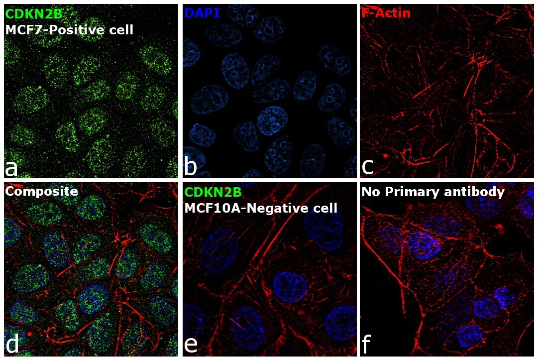
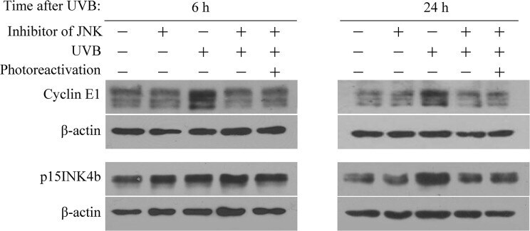
- Fig 3 Photorepair of CPDs prevents altered expression of cyclin E1 and p15INK4b protein in UVB irradiated HaCaT cells. Cells were transfected with lipofectamine-complexed CPD-PL Psi-mRNA, 12 hs later irradiated with 20 mJ/cm 2 UVB and immediately exposed to photoreactivating light (active CPD-photolyase) or kept in the dark (inactive CPD-photolyase) for 1 h. Subsequently, cells were cultured at 37degC until harvested at the indicated time after UVB irradiation. (A) The expression of cyclin E1 and p15INK4b were analyzed by Western blot. (B) Quantitation of western blots displays relative changes in protein expression normalized to beta-actin. Pixel densities were calculated relative to those obtained with cells that were not UVB irradiated (pecked lines). Significance was assessed by two-tailed, unpaired t -test (asterisk, p
- Submitted by
- Invitrogen Antibodies (provider)
- Main image
- Experimental details
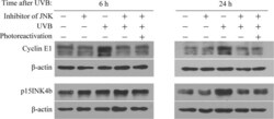
- Fig 4 Induction of cyclin E1 and p15INK4b protein expression upon UVB exposure is regulated through the JNK signalling pathway in HaCaT cells. Keratinocytes transfected with CPD-PL Psi-mRNA were incubated in serum-free medium supplemented with JNK inhibitor (SP600125) for 1 h. Immediately thereafter, cells were irradiated with a physiological dose of UVB or left untreated followed by exposure to photoreactivating light (or not) for 1 h. The cells were cultured further in serum-free medium supplemented with the inhibitor. Cells were harvested for western blot assay at the indicated time. Protein levels of cyclin E1, p15INK4b, and beta-actin are noted. The figure shows representative results from three independent experiments.
- Submitted by
- Invitrogen Antibodies (provider)
- Main image

- Experimental details
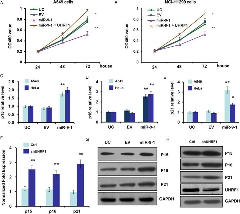
- Figure 5. Suppression of UHRF1 by miR-9 to 1 or shRNA causes expression upregulation of p15, p16, p21 in A549 cells. (A, B) Growth curves of miR-9 to 1, UHRF1, and EV-infected A549 (A) and NCI-H1299 (B) cells were conducted by CCK-8 assay. The OD value at 450 nm represented the viable cell numbers. (C-E) Quantitative PCR results showed the mRNA levels of p15 (C), p16 (D), and p21 (E) were induced after miR-9 to 1stable transfection of A549 cells. (F) Quantitative PCR arrays for the mRNA levels of p15 (C), p16 (D), and p21 (E) were induced after UHRF1 RNAi in A549 cells. (G, H) Western blot to quantify protein levels of p15, p16, and p21 after miR-9 to 1 stable transfection (G or UHRF1 RNAi [H] in A549 cells). Abbreviations: miR-9 to 1, microRNA-9 to 1; PCR, polymerase chain reaction; UHRF1, ubiquitin-like containing PHD and RING finger domains 1.
- Submitted by
- Invitrogen Antibodies (provider)
- Main image
- Experimental details
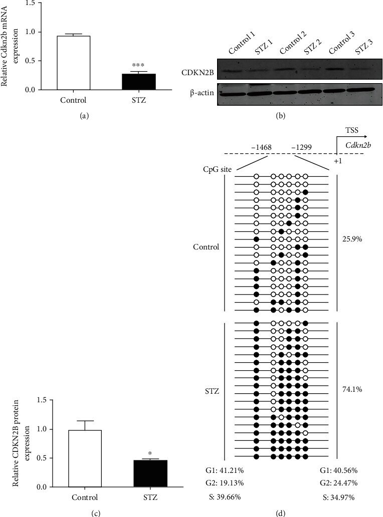
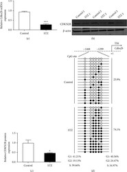
- Figure 4 DNA hypermethylation suppressed CDKN2B expression during diabetic retinopathy. (a) Quantitative analysis of CDKN2B mRNA expression in the control and STZ groups. (b) Western blot analysis was used to assess the protein levels of CDKN2B in the control and STZ groups. (c) Western blot analysis indicated the significantly decreased CDKN2B protein expression in the STZ group. (d) The position of the CpG sites at the upstream regulatory genomic locus of CDKN2B. Analysis of DNA methylation status at the upstream regulatory locus of the CDKN2B gene in control and WH groups determined by BSP. TSS: transcriptional start site. * P < 0.05, *** P < 0.001.