 Explore
 Validate
 Learn
Learn Western blot
Antibody data
- Antibody Data
- Antigen structure
- References [1]
- Comments [0]
- Validations
- Western blot [9]
- Immunocytochemistry [1]
- Immunohistochemistry [1]
- Other assay [1]
Submit
Validation data
Reference
Comment
Report error
- Product number
- PA5-23219 - Provider product page

- Provider
- Invitrogen Antibodies
- Product name
- TGM2 Polyclonal Antibody
- Antibody type
- Polyclonal
- Antigen
- Other
- Reactivity
- Human
- Host
- Rabbit
- Isotype
- IgG
- Vial size
- 100 µg
- Concentration
- 1 mg/mL
- Storage
- Store at 4°C short term. For long term storage, store at -20°C, avoiding freeze/thaw cycles.
Submitted references Intracellular construction of topology-controlled polypeptide nanostructures with diverse biological functions.
Li LL, Qiao SL, Liu WJ, Ma Y, Wan D, Pan J, Wang H
Nature communications 2017 Nov 2;8(1):1276
Nature communications 2017 Nov 2;8(1):1276
No comments: Submit comment
Supportive validation
- Submitted by
- Invitrogen Antibodies (provider)
- Main image
- Experimental details
- Western blot analysis of TGM2 using a polyclonal antibody (Product # PA5-23219).
- Submitted by
- Invitrogen Antibodies (provider)
- Main image

- Experimental details
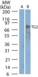
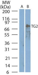
- Western blot analysis of TGM2 in A) BxPC-3 cell lysate; B) MCF-7 negative control cell lysate. Samples were incubated in TGM2 polyclonal antibody (Product # PA5-23219 using a dilution of 2.0 µg/mL.
- Submitted by
- Invitrogen Antibodies (provider)
- Main image
- Experimental details
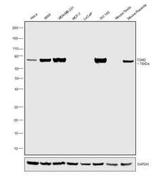
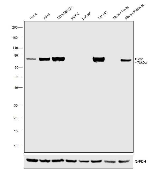
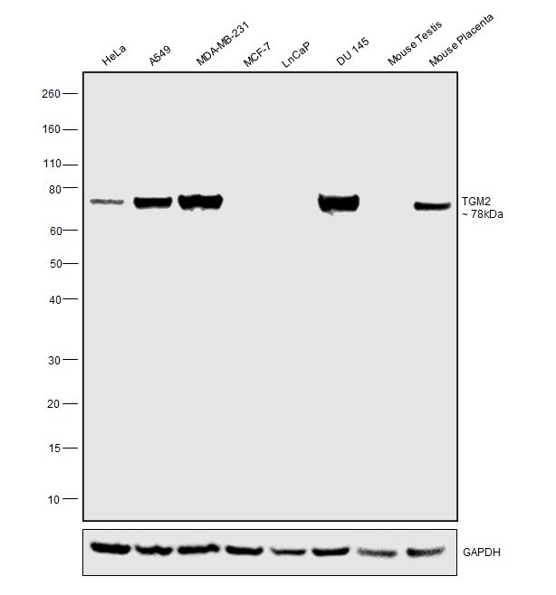
- Western blot was performed using Anti-TGM2 Polyclonal Antibody(Product # PA5-23219) and a 78kDa band corresponding to TGM2 was observed across cell lines and tissue tested. Whole cell extracts (30 µg lysate) of HeLa (Lane 1), A549 (Lane 2), MDA-MB-231 (Lane 3), MCF7 (Lane 4), LNCaP (Lane 5) and DU 145 (Lane 6).Tissue extracts of Mouse Testis (Lane 7) and Mouse Placenta (Lane 8), were electrophoresed using NuPAGE™ 4-12% Bis-Tris Protein Gel (Product # NP0321BOX). Resolved proteins were then transferred onto a Nitrocellulose membrane (Product # IB23001) by iBlot® 2 Dry Blotting System (Product # IB21001). The blot was probed with the primary antibody (2ug/ml) and detected by chemiluminescence with Goat anti-Rabbit IgG (H+L) Superclonal™ Secondary Antibody, HRP (Product # A27036, 1:4000 dilution) using the iBright FL 1000 (Product # A32752). Chemiluminescent detection was performed using Novex® ECL Chemiluminescent Substrate Reagent Kit (Product # WP20005).
- Submitted by
- Invitrogen Antibodies (provider)
- Main image

- Experimental details
- Western blot analysis of TGM2 in A) BxPC-3 cell lysate; B) MCF-7 negative control cell lysate. Samples were incubated in TGM2 polyclonal antibody (Product # PA5-23219 using a dilution of 2.0 µg/mL.

- Submitted by
- Invitrogen Antibodies (provider)
- Main image

- Experimental details
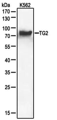
- Western blot analysis of TGM2 in extracts from K562 cells. Samples were incubated in TGM2 polyclonal antibody (Product # PA5-23219) using a dilution of 1:100.
- Submitted by
- Invitrogen Antibodies (provider)
- Main image

- Experimental details
- Western blot analysis of TGM2 in 0.5 mg/mL MCF-7 lysate. Samples were incubated in TGM2 polyclonal antibody (Product # PA5-23219). This experiment was performed under reducing conditions using the 12-230 kDa separation system.

- Submitted by
- Invitrogen Antibodies (provider)
- Main image
- Experimental details
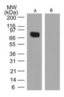
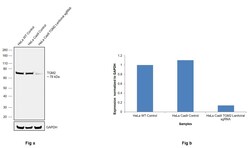
- CRISPR-Cas9 mediated genome editing ofTGM2 (as confirmed by next generation sequencing) was achieved by using LentiArray™ Lentiviral sgRNA (Product # A32042, Assay ID CRISPR979226_LV) and LentiArray Cas9 Lentivirus (Product # A32064). Fig (a) Western blot analysis of TGM2 was performed by loading 50 µg of HeLa Wild type (Lane 1), HeLa Cas9 (Lane 2) and HeLa Cas9 cells transduced with TGM2 Lentiviral sgRNA (Lane 3) whole cell extracts. The samples were electrophoresed using NuPAGE™ Novex™ 4-12% Bis-Tris Protein Gel (Product # NP0322BOX). Resolved proteins were then transferred onto a nitrocellulose membrane (Product # IB23001) by iBlot® 2 Dry Blotting System (Product # IB21001). The blot was probed with Anti-TGM2 Polyclonal Antibody (Product # PA5-23219) using 2 µg/mL dilution and Goat anti-Rabbit IgG (H+L) Superclonal™ Recombinant Secondary Antibody, HRP (Product # A27036 1:5,000 dilution). Chemiluminescent detection was performed using Novex® ECL Chemiluminescent Substrate Reagent Kit (Product # WP20005). A reduced signal in sgRNA transduced cells using the LentiArray™ CRISPR product line confirms that antibody is specific toTGM2 (Fig (b)).

- Submitted by
- Invitrogen Antibodies (provider)
- Main image

- Experimental details
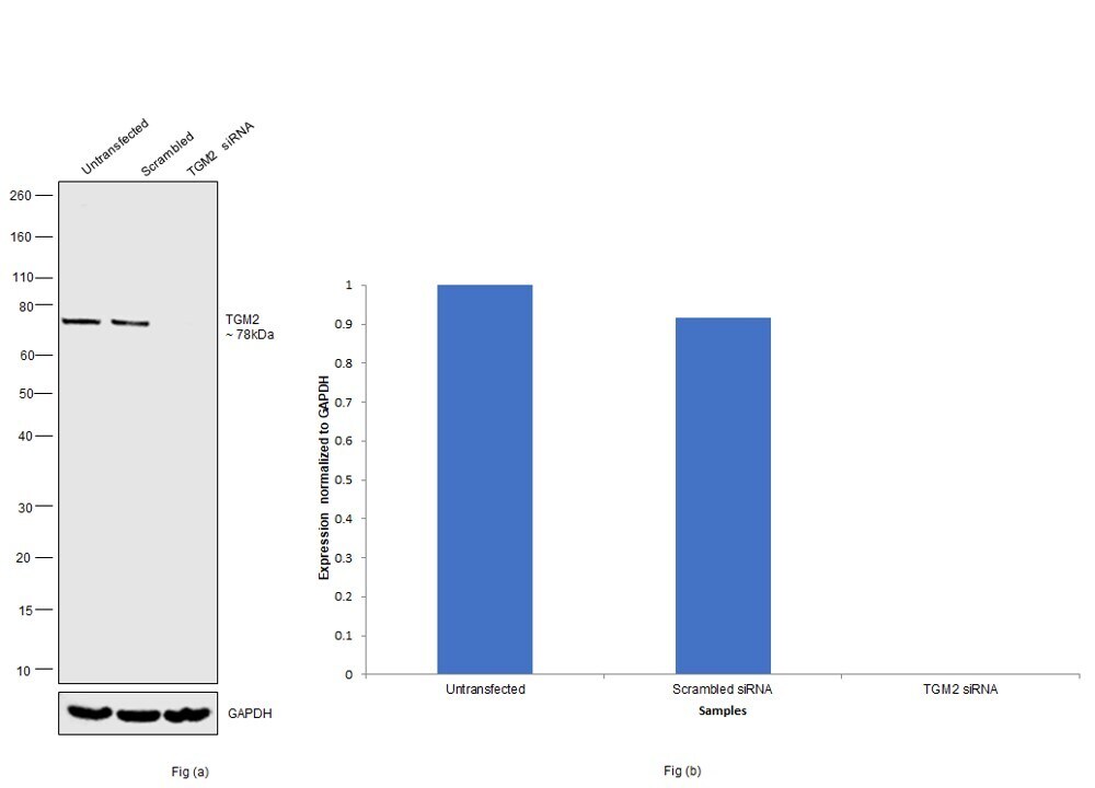
- Knockdown of TGM2 was achieved by transfecting HeLa with TGM2 specific siRNAs (Silencer® select Product # S14087, S14088). Western blot analysis (Fig. a) was performed using Whole cell extracts from the TGM2 knockdown cells (lane 3), non-targeting scrambled siRNA transfected cells (lane 2) and untransfected cells (lane 1). The blot was probed with TGM2 Polyclonal Antibody (Product # PA5-23219, 2ug/ml ) and Goat anti-Rabbit IgG (H+L) Superclonal™ Recombinant Secondary Antibody, HRP (Product # A27036, 1:4000 dilution). Densitometric analysis of this western blot is shown in histogram (Fig. b). Decrease in signal upon siRNA mediated knock down confirms that antibody is specific to TGM2.
- Submitted by
- Invitrogen Antibodies (provider)
- Main image

- Experimental details
- Western blot was performed using Anti-TGM2 Polyclonal Antibody(Product # PA5-23219) and a 78kDa band corresponding to TGM2 was observed across cell lines and tissue tested. Whole cell extracts (30 µg lysate) of HeLa (Lane 1), A549 (Lane 2), MDA-MB-231 (Lane 3), MCF7 (Lane 4), LNCaP (Lane 5) and DU 145 (Lane 6).Tissue extracts of Mouse Testis (Lane 7) and Mouse Placenta (Lane 8), were electrophoresed using NuPAGE™ 4-12% Bis-Tris Protein Gel (Product # NP0321BOX). Resolved proteins were then transferred onto a Nitrocellulose membrane (Product # IB23001) by iBlot® 2 Dry Blotting System (Product # IB21001). The blot was probed with the primary antibody (2ug/ml) and detected by chemiluminescence with Goat anti-Rabbit IgG (H+L) Superclonal™ Secondary Antibody, HRP (Product # A27036, 1:4000 dilution) using the iBright FL 1000 (Product # A32752). Chemiluminescent detection was performed using Novex® ECL Chemiluminescent Substrate Reagent Kit (Product # WP20005).
Supportive validation
- Submitted by
- Invitrogen Antibodies (provider)
- Main image
- Experimental details
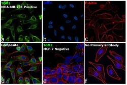
- Immunofluorescence analysis of TGM2 was performed using 70% confluent log phase MDA-MB-231 and MCF7 cells. The cells were fixed with 4% Paraformaldehyde for 10 minutes, permeabilized with 0.1% Triton™ X-100 for 10 minutes, and blocked with 2% BSA for 10 minutes at room temperature. The cells were labeled with TGM2 Polyclonal Antibody (Product # PA5-23219) at 5 µg/mL in 0.1% BSA, incubated at 4 degree celsius overnight and then labeled with Alexa Fluor Plus 488 donkey anti-rabbit IgG secondary antibody -A32790), (1:2,000 dilution) for 45 minutes at room temperature (Panel a: Green). Nuclei (Panel b: Blue) were stained with ProLong™ Diamond Antifade Mountant with DAPI (Product # P36962). F-actin (Panel c: Red) was stained with Rhodamine Phalloidin (Product # R415, 1:300 dilution). Panel d represents the merged image showing cytoplasmic localization. Panel e represents MCF7 cells having no expression of TGM2. Panel f represents control cells with no primary antibody to assess background. The images were captured at 60X magnification.

Supportive validation
- Submitted by
- Invitrogen Antibodies (provider)
- Main image

- Experimental details
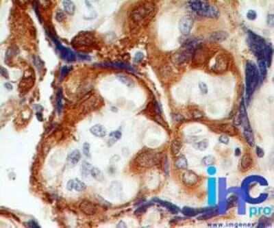
- Immunohistochemical analysis of TGM2 in formalin-fixed, paraffin-embedded human placenta. Samples were incubated in TGM2 polyclonal antibody (Product # PA5-23219) using a dilution of 1.0 µg/mL. Note cytoplasmic staining of trophoblastic cells.
Supportive validation
- Submitted by
- Invitrogen Antibodies (provider)
- Main image
- Experimental details
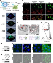
- Fig. 3 Intracellular polymerization and in situ aggregation of peptide monomers. a TG2-catalyzed intracellular polymerization of peptide monomers was monitored by fluorescent resonance energy transfer (FRET) technique. The 3D confocal images displayed the FRET effect of control, P4 , P7 , and P9 inside HeLa cells at 12 h-post treatment. The feed ratio of FITC-peptide (fluorescein isothiocyannate-peptide) and CO-peptide (coumarin-peptide) was 1:1. b Confocal images displayed the nanostructures (green) formed from P4 (at 4 degC) and ( P7 , P9 ) (at 37 degC). P8 , P4 , P7 , and P9 were ELP random coil, UCST-type ELP, LCST-type ELP, and ELP gel, respectively. Cell membrane, red. Scale bar, 30 mum. c The ultrathin cell sections of HeLa cells. Fe-chelated P 18 was used to identify the formed nanostructures and for contrast enhancement. Red arrows indicated P 18 -P4 3D nanoparticles; red dotted circle indicated P 18 -P9 gel. Scale bar, 1 mum. d Energy dispersive spectrometry (EDS) of the nanostructures formed from P 18 -P4 . The Fe element signal (red highlighted) made the P 18 -P4 from the biological background stand out. e MCF-7 and HeLa cells were lysed. TG2 protein was examined by western blotting. f Fluorescein isothiocyannate (FITC)-labeled P7 and P9 were incubated with MCF-7 or HeLa cells for fluorescence imaging. FITC-P7 and FITC-P9 , green; nucleus, blue. Scale bar, 30 mum. g The expression of TG2 protein in SH-SY5Y cells was modulated by normoxia (N) and hypoxia (H) condit
