Explore
 Validate
 Learn
Learn Western blot
Antibody data
- Antibody Data
- Antigen structure
- References [0]
- Comments [0]
- Validations
- Western blot [5]
- Immunocytochemistry [2]
- Immunohistochemistry [1]
Submit
Validation data
Reference
Comment
Report error
- Product number
- PA5-20754 - Provider product page

- Provider
- Invitrogen Antibodies
- Product name
- Claudin 1 Polyclonal Antibody
- Antibody type
- Polyclonal
- Antigen
- Synthetic peptide
- Description
- A suggested positive control is HepG2 cell lysate.
- Concentration
- 1 mg/mL
No comments: Submit comment
Supportive validation
- Submitted by
- Invitrogen Antibodies (provider)
- Main image
- Experimental details
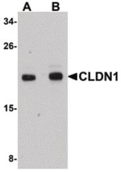
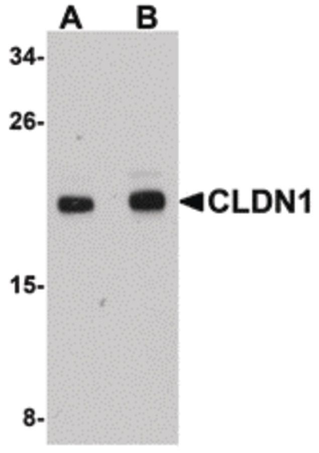
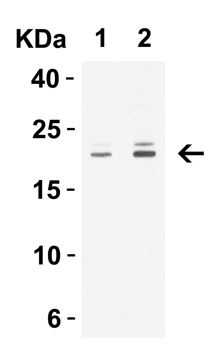
- Western blot analysis of HepG2 cell lysate using a CLDN1 polyclonal antibody (Product # PA5-20754) at (A) 1 and (B) 2 µg/mL.
- Submitted by
- Invitrogen Antibodies (provider)
- Main image

- Experimental details
- Western Blot Validation in Human HepG2 Cell Lysate. Loading: 15 µg of lysates per lane. Antibodies: Claudin 1 Polyclonal Antibody (Product # PA5-20754) (A: 1 µg/mL and B: 2 µg/mL), 1h incubation at RT in 0.05 NFDM/TBST. Secondary: Goat anti-rabbit IgG HRP conjugate at 1:10,000 dilution.

- Submitted by
- Invitrogen Antibodies (provider)
- Main image
- Experimental details
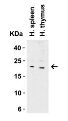
- Western Blot Validation in Human Tissue Lysates. Loading: 15 µg of lysates per lane. Antibodies: Claudin 1 Polyclonal Antibody (Product # PA5-20754) (1 µg/mL) , 1h incubation at RT in 0.05 NFDM/TBST. Secondary: Goat anti-rabbit IgG HRP conjugate at 1:10,000 dilution.

- Submitted by
- Invitrogen Antibodies (provider)
- Main image

- Experimental details
- Western Blot Validation in Human Tissue Lysates. Loading: 15 µg of lysates per lane. Antibodies: Claudin 1 Polyclonal Antibody (Product # PA5-20754) (1 µg/mL) , 1h incubation at RT in 0.05 NFDM/TBST. Secondary: Goat anti-rabbit IgG HRP conjugate at 1:10,000 dilution.
- Submitted by
- Invitrogen Antibodies (provider)
- Main image
- Experimental details
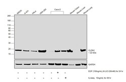
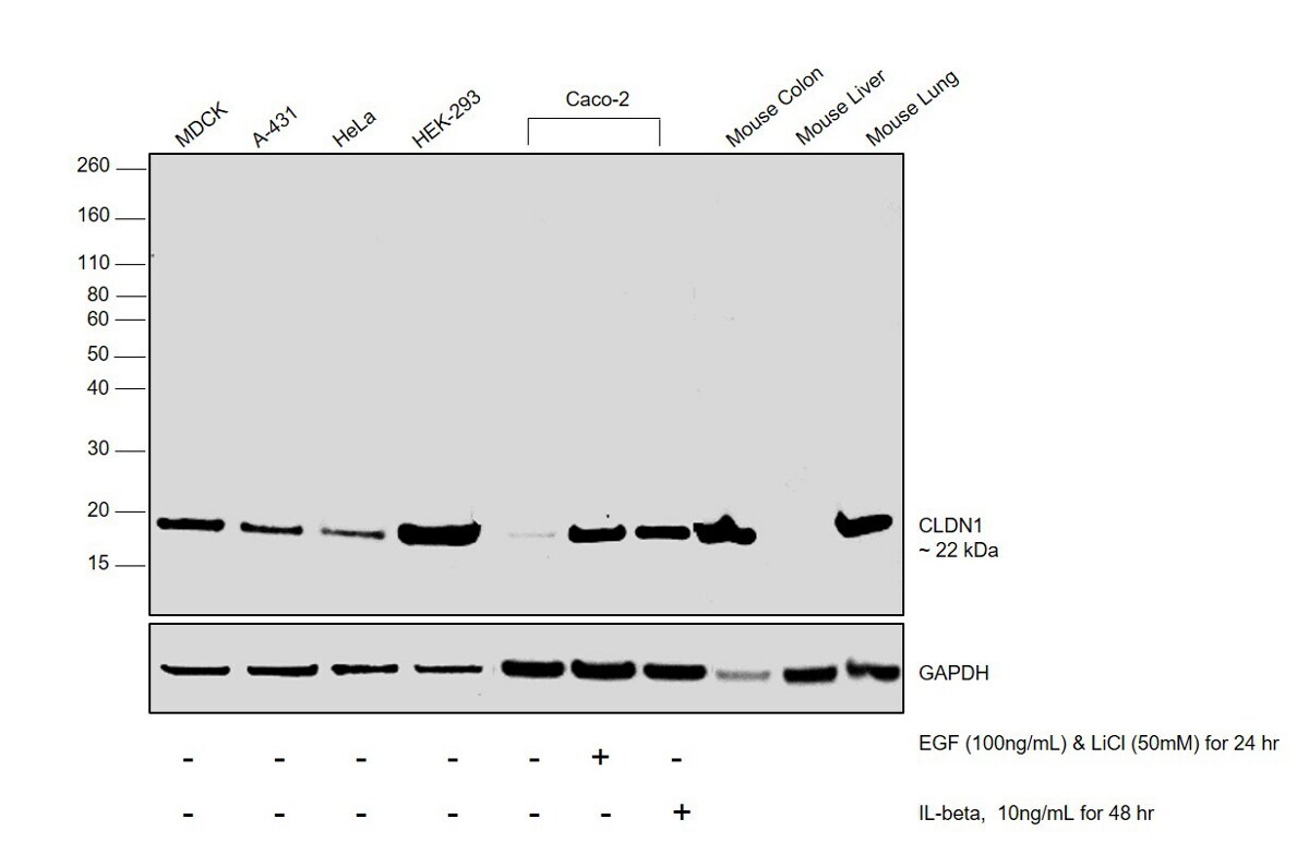
- Western blot was performed using Anti-CLDN1 Polyclonal Antibody (Product # PA5-20754) and a 22 kDa band corresponding to CLDN1 was observed across cell lines tested and increased upon EGF, LiCl treatment and IL-beta treatment. Membrane enriched extracts (30 µg lysate) of MDCK (Lane 1), A-431 (Lane 2), HeLa (Lane 3), HEK-293 (Lane 4) Caco-2 (Lane 5), Caco-2 treated with EGF (100ng/mL) and LiCl (50mM) simultaneously for 24 Hours (Lane 6), Caco-2 treated with IL-beta (10ng/mL for 48 Hours) (Lane 7), tissue extracts (30ug lysate) of Mouse Kidney (Lane 8), Mouse Liver (Lane 9) and Mouse Lung (Lane 10) were electrophoresed using Novex® NuPAGE® 12 % Bis-Tris gel (Product # NP0342BOX). Resolved proteins were then transferred onto a nitrocellulose membrane (Product # IB23001) by iBlot® 2 Dry Blotting System (Product # IB21001). The blot was probed with the primary antibody (1µg/ml) and detected by chemiluminescence with Goat anti-Rabbit IgG (H+L), Superclonal™ Recombinant Secondary Antibody, HRP (Product # A27036, 1:4000 dilution) using the iBright FL 1000 (Product # A32752). Chemiluminescent detection was performed using Novex® ECL Chemiluminescent Substrate Reagent Kit (Product # WP20005).
Supportive validation
- Submitted by
- Invitrogen Antibodies (provider)
- Main image
- Experimental details
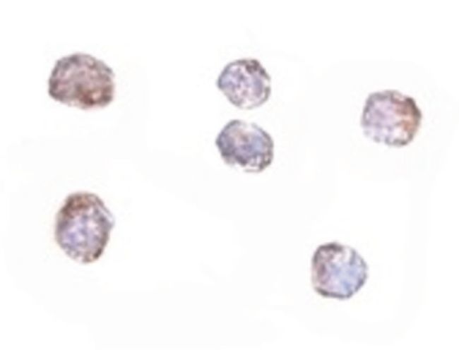
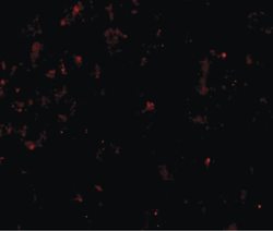
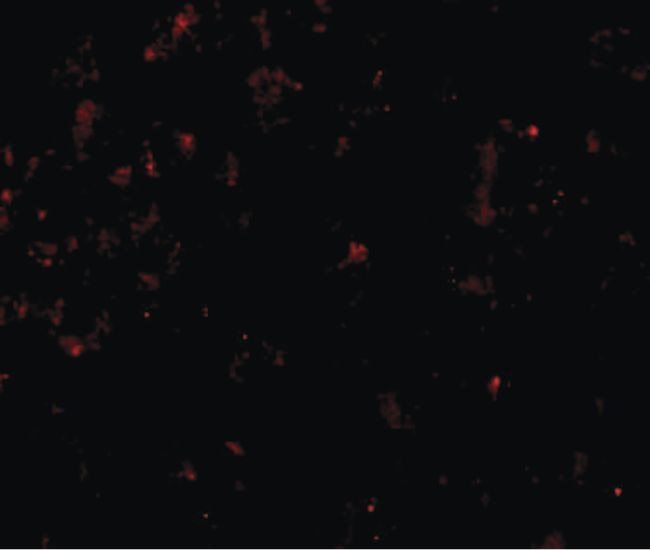
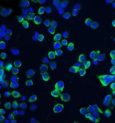
- Immunofluorescent analysis of HepG2 cells using a CLDN1 polyclonal antibody (Product # PA5-20754) at a 20 µg/mL dilution.
- Submitted by
- Invitrogen Antibodies (provider)
- Main image
- Experimental details
- Immunofluorescent analysis of 4% paraformaldehyde-fixed HepG2 cells labeling CLDN1 with Claudin 1 Polyclonal Antibody (Product # PA5-20754) at 20 µg/mL, followed by goat anti-rabbit IgG secondary antibody at 1:500 dilution (green) and DAPI staining (blue).

Supportive validation
- Submitted by
- Invitrogen Antibodies (provider)
- Main image
- Experimental details
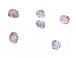
- Immunocytochemistry staining of HepG2 cells using a CLDN1 polyclonal antibody (Product # PA5-20754) at a 5 µg/mL dilution.