Explore
 Validate
 Learn
Learn Western blot
Antibody data
- Antibody Data
- Antigen structure
- References [0]
- Comments [0]
- Validations
- Western blot [2]
- Immunocytochemistry [2]
- Immunohistochemistry [1]
Submit
Validation data
Reference
Comment
Report error
- Product number
- PA5-32350 - Provider product page

- Provider
- Invitrogen Antibodies
- Product name
- Claudin 1 Polyclonal Antibody
- Antibody type
- Polyclonal
- Antigen
- Synthetic peptide
- Description
- Heat-mediated antigen retrieval is recommended prior to staining, using a 10mM citrate buffer, pH 6.0, for 10 minutes followed by cooling at room temperature for 20 min. Following antigen retrieval, incubate samples with primary antibody for 10 min at room temperature. A suggested positive control is skin, breast carcinoma or ovarian carcinoma.
- Reactivity
- Human, Mouse, Rat
- Host
- Rabbit
- Isotype
- IgG
- Vial size
- 500 µL
- Storage
- Store at 4°C short term. For long term storage, store at -20°C, avoiding freeze/thaw cycles.
No comments: Submit comment
Supportive validation
- Submitted by
- Invitrogen Antibodies (provider)
- Main image

- Experimental details
- Western blot analysis of Hela Cells using anti-Claudin-1 Polyclonal Antibody (Product # PA5-32350). The recommened dilution for this antibody in western blot applications is 1:25.

- Submitted by
- Invitrogen Antibodies (provider)
- Main image
- Experimental details
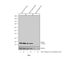
- Knockout of CLDN1 was achieved by CRISPR-Cas9 genome editing using LentiArray™ Lentiviral sgRNA (Product # A32042, Assay ID CRISPR961566_LV) and LentiArray Cas9 Lentivirus (Product # A32064). Western blot analysis of CLDN1 was performed by loading 30 µg of Caco-2 wild type (Lane 1), Caco-2 wild type treated with 100 ng/mL EGF and 50mM LiCi for 24hrs (Lane 2),Caco-2 Cas9 (Lane 3), Caco-2 Cas9 treated with 100 ng/mL EGF and 50mM LiCi for 24hrs (Lane 4), Caco-2 CLDN1 KO (Lane 5) and Caco-2 CLDN1 KO treated with 100 ng/mL EGF and 50mM LiCi for 24hrs (Lane 6) membrane enriched extracts. The samples were electrophoresed using NuPAGE™ Novex™ 4-12% Bis-Tris Protein Gel (Product # NP0322BOX). Resolved proteins were then transferred onto a nitrocellulose membrane (Product # IB23001) by iBlot® 2 Dry Blotting System (Product # IB21001). The blot was probed with Anti-Claudin 1 Polyclonal Antibody (Product # PA5-32350, 1:50 dilution) and Goat anti-Rabbit IgG (H+L) Superclonal™ Recombinant Secondary Antibody, HRP (Product # A27036, 1:5000 dilution) using the iBright FL 1000 (Product # A32752). Chemiluminescent detection was performed using Novex® ECL Chemiluminescent Substrate Reagent Kit (Product # WP20005). Loss of signal upon CRISPR mediated knockout (KO) using the LentiArray™ CRISPR product line confirms that antibody is specific to CLDN1.

Supportive validation
- Submitted by
- Invitrogen Antibodies (provider)
- Main image

- Experimental details
- Immunofluorescence analysis of Claudin 1 was performed using 90% confluent log phase Caco-2 cells. The cells were fixed with 4% paraformaldehyde for 10 minutes, permeabilized with 0.1% Triton™ X-100 for 15 minutes, and blocked with 1% BSA for 1 hour at room temperature. The cells were labeled with Claudin 1 Rabbit Polyclonal Antibody(Product # PA5-32350) at 1:100 dilution in 0.1% BSA, incubated at 4 degree Celsius overnight and then labeled with Goat anti-Rabbit IgG (H+L) Superclonal™ Secondary Antibody, Alexa Fluor® 488 conjugate (Product # A27034) at a dilution of 1:2000 for 45 minutes at room temperature (Panel a: green). Nuclei (Panel b: blue) were stained with SlowFade® Gold Antifade Mountant with DAPI (Product # S36938). F-actin (Panel c: red) was stained with Rhodamine Phalloidin (Product # R415, 1:300). Panel d represents the merged image showing localization at the cell junction. Panel e represents control cells with no primary antibody to assess background. The images were captured at 60X magnification.

- Submitted by
- Invitrogen Antibodies (provider)
- Main image

- Experimental details
- Knockdown of Claudin 1 was achieved by transfecting Caco-2 cells with Claudin 1 specific siRNA (Silencer® select Product # s17315, s17316). Immunofluorescence analysis was performed using untransfected Caco-2 cells (panels a, d), transfected with non-specific scrambled siRNA (panels b,e) and transfected with Claudin 1 specific siRNAs (panel c,f). Cells were fixed, permeabilized, and probed with Claudin 1 Polyclonal Antibody (Product # PA5-32350, 1:250 dilution), followed by labelling with Goat anti-Mouse IgG (H+L) Superclonal™ Secondary Antibody, Alexa Fluor 488 (Product # A27034, 1:2000). Nuclei (blue) were stained using ProLong™ Diamond Antifade Mountant with DAPI (Product # P36962) and Rhodamine Phalloidin (Product # R415, 1:300) was used for cytoskeletal F-actin (red) staining. Reduction of specific junctional localization was observed upon siRNA mediated knockdown (panel c,f) confirming specificity of the antibody to Claudin 1. The images were captured at 60X magnification.

Supportive validation
- Submitted by
- Invitrogen Antibodies (provider)
- Main image

- Experimental details
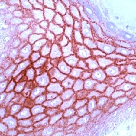
- Immunohistochemical analysis of Claudin-1 using anti-Claudin-1 Polyclonal Antibody (Product # PA5-32350) in Skin Cancer Tissue. The recommened dilution for this antibody in immunohistochemistry applications is 1:200.