Explore
 Validate
 Learn
Learn Western blot
Antibody data
- Antibody Data
- Antigen structure
- References [2]
- Comments [0]
- Validations
- Western blot [2]
- Immunocytochemistry [1]
- Immunoprecipitation [2]
- Immunohistochemistry [2]
Submit
Validation data
Reference
Comment
Report error
- Product number
- GTX105202 - Provider product page

- Provider
- GeneTex
- Proper citation
- GeneTex Cat#GTX105202, RRID:AB_11173475
- Product name
- OCT1 antibody
- Antibody type
- Polyclonal
- Reactivity
- Human, Rat
- Host
- Rabbit
Submitted references Long non-coding RNA TUG1 contributes to tumorigenesis of human osteosarcoma by sponging miR-9-5p and regulating POU2F1 expression.
Bioinformatics Data Mining Approach Suggests Coexpression of AGTPBP1 with an ALS-linked Gene C9orf72.
Xie CH, Cao YM, Huang Y, Shi QW, Guo JH, Fan ZW, Li JG, Chen BW, Wu BY
Tumour biology : the journal of the International Society for Oncodevelopmental Biology and Medicine 2016 Nov;37(11):15031-15041
Tumour biology : the journal of the International Society for Oncodevelopmental Biology and Medicine 2016 Nov;37(11):15031-15041
Bioinformatics Data Mining Approach Suggests Coexpression of AGTPBP1 with an ALS-linked Gene C9orf72.
Kitano S, Kino Y, Yamamoto Y, Takitani M, Miyoshi J, Ishida T, Saito Y, Arima K, Satoh J
Journal of central nervous system disease 2015;7:15-26
Journal of central nervous system disease 2015;7:15-26
No comments: Submit comment
Supportive validation
- Submitted by
- GeneTex (provider)
- Main image
- Experimental details
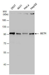
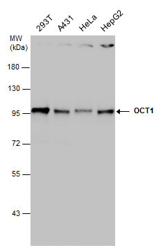
- Sample (30 ug of whole cell lysate) A: 293T B: A431 C: HeLa D: HepG2 7.5% SDS PAGE GTX105202 diluted at 1:1000

- Submitted by
- GeneTex (provider)
- Main image
- Experimental details
- Various whole cell extracts (30 £gg) were separated by 7.5% SDS-PAGE, and the membrane was blotted with OCT1 antibody (GTX105202) diluted at 1:1000.
Supportive validation
- Submitted by
- GeneTex (provider)
- Main image
- Experimental details
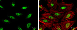
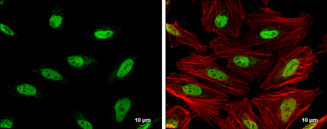
- OCT1 antibody detects OCT1 protein at nucleus by immunofluorescent analysis.Sample: HeLa cells were fixed in 4% paraformaldehyde at RT for 15 min.Green: OCT1 protein stained by OCT1 antibody (GTX105202) diluted at 1:500.Red: phalloidin, a cytoskeleton marker, stained by () diluted at 1:200.Blue: Hoechst 33342 staining.Scale bar = 10 £gm.
Supportive validation
- Submitted by
- GeneTex (provider)
- Main image

- Experimental details
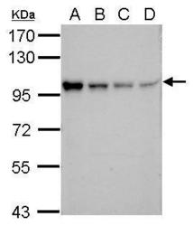
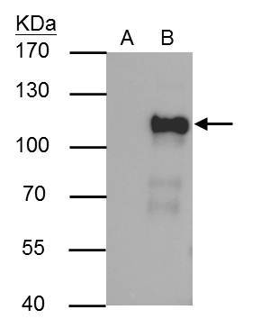
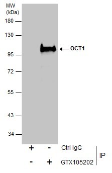
- OCT1 antibody immunoprecipitates OCT1 protein in IP experiments. IP Sample: 293T whole cell lysate/extract A : Control with 2.5 £gg of pre-immune rabbit IgG B : Immunoprecipitation of OCT1 by 2.5 £gg of OCT1 antibody (GTX105202) 7.5% SDS-PAGE The immunoprecipitated OCT1 protein was detected by OCT1 antibody (GTX105202) diluted at 1 : 1000. EasyBlot anti-rabbit IgG (HRP) (GTX221666-01) was used as a secondary reagent.
- Submitted by
- GeneTex (provider)
- Main image
- Experimental details
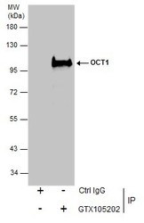
- Immunoprecipitation of OCT1 protein from 293T whole cell extracts using 5 £gg of OCT1 antibody (GTX105202).Western blot analysis was performed using OCT1 antibody (GTX105202).EasyBlot anti-Rabbit IgG (GTX221666-01) was used as a secondary reagent.
Supportive validation
- Submitted by
- GeneTex (provider)
- Main image
- Experimental details
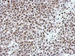
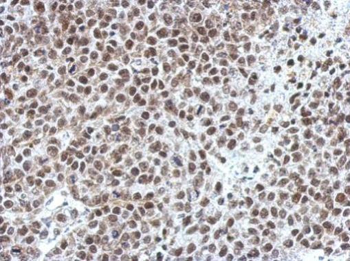
- Immunohistochemical analysis of paraffin-embedded Hela xenograft, using OCT1(GTX105202) antibody at 1:750 dilution.
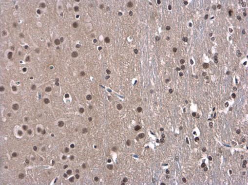
- Submitted by
- GeneTex (provider)
- Main image
- Experimental details
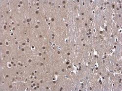
- OCT1 antibody detects OCT1 protein at nucleus in rat brain by immunohistochemical analysis. Sample: Paraffin-embedded rat brain. OCT1 antibody (GTX105202) diluted at 1:500.