Explore
 Validate
 Learn
Learn Western blot
Antibody data
- Antibody Data
- Antigen structure
- References [0]
- Comments [0]
- Validations
- Western blot [1]
- Immunocytochemistry [1]
Submit
Validation data
Reference
Comment
Report error
- Product number
- 14-9736-82 - Provider product page

- Provider
- Invitrogen Antibodies
- Product name
- OCT1 (POU2F1) Monoclonal Antibody (YL15), eBioscience™
- Antibody type
- Monoclonal
- Antigen
- Recombinant full-length protein
- Description
- Description: This YL15 monoclonal antibody recognizes human OCT1 (POU2F1) and is predicted to recognize mouse POU2F1.
- Antibody clone number
- YL15
- Concentration
- 0.5 mg/mL
No comments: Submit comment
Supportive validation
- Submitted by
- Invitrogen Antibodies (provider)
- Main image

- Experimental details
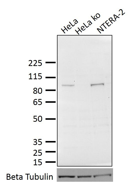
- Western blot analysis was performed on whole cell extracts of untreated HeLa cells (lane 1), CRISPR-Cas9 modified HeLa cells (lane 2) and NTERA-2 cells (lane 3). The proteins were separated on a 4-12% Nupage Bis-Tris Gel (Product # NP0336BOX) using MOPS SDS Running Buffer (Product # NP0001-02). Protein transfer to a PVDF membrane (Product # IB24002) was achieved using the iBlot 2 system. Subsequently, the membrane was blocked with 5% milk and probed overnight at 4 C with 4.0 µg/mL of POU2F1 Monoclonal Antibody, Unconjugated, followed by anti-Mouse IgG, HRP secondary antibody. Protein detection was performed using SuperSignal West Pico PLUS Chemiluminescence Substrate (Product # 34580) and the Invitrogen iBright FL1000 Imaging System. As expected, Clone YL15 detects the ~95 kDa band corresponding to the POU2F1 protein in lanes 1 and 3 but not in lane 2. Protein size was determined using Spectra™ Multicolor Broad Range Protein Ladder (Product # 26634). Beta Tubulin Monoclonal Anitbody, Alexa Fluor 488 (Product # MA5-16308-A488) was used as a loading control.
Supportive validation
- Submitted by
- Invitrogen Antibodies (provider)
- Main image

- Experimental details
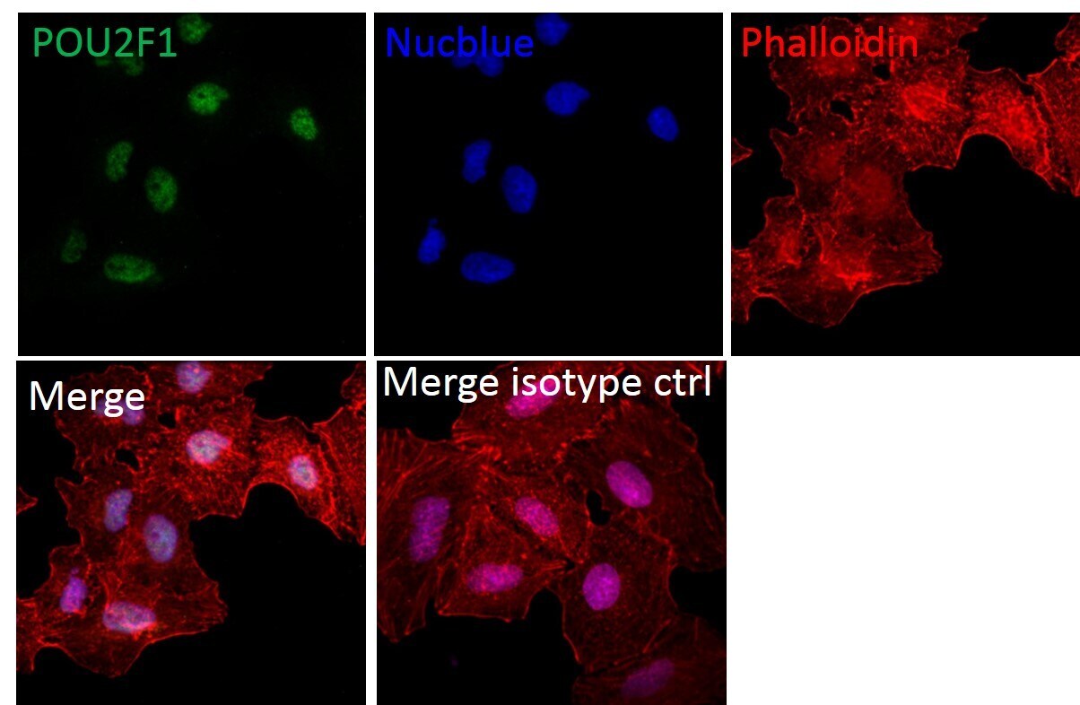
- Immunofluorescent analysis of POU2F1 (green) in A549 cells (60 % confluency). The cells were fixed with 4% paraformaldehyde for 15 minutes at room temperature, permeabilized with 0.1% Triton X-100 for 10 minutes at 37 C, and blocked with 10% Normal Donkey Serum in PBS for 1 hour at room temperature. Cells were then stained with OCT1 (POU2F1) Monoclonal Antibody, Unconjugated at 1.25 µg/mL Mouse IgG1 isotype control at room temperature in blocking buffer, followed by Donkey anti-Mouse IgG (H+L) Alexa Fluor Plus 488 (Product # A32766) at 1:5000 in blocking buffer for 1 hour at room temperature. Cell nuclei were stained with NucBlueTM stain in ProLong™ Glass Antifade Mount (Product # P36985) (blue). The cytoskeleton was visualized using Rhodamine Phalloidin (red) (Product # R415). Images were acquired at 40x magnification with a Zeiss LSM 800 confocal. The upper panel shows cells stained with clone YL15. No staining was observed with mouse IgG1 isotype control (lower panel, Merged isotype).