 Explore
 Validate
 Learn
Learn Western blot
Antibody data
- Antibody Data
- Antigen structure
- References [1]
- Comments [0]
- Validations
- Western blot [2]
- Immunocytochemistry [2]
Submit
Validation data
Reference
Comment
Report error
- Product number
- 710221 - Provider product page

- Provider
- Invitrogen Antibodies
- Product name
- Claudin 2 Recombinant Polyclonal Antibody (3HCLC)
- Antibody type
- Polyclonal
- Antigen
- Synthetic peptide
- Description
- This antibody is predicted to react with equine, mouse and rat based on sequence homology.
- Antibody clone number
- 3HCLC
- Concentration
- 0.5 mg/mL
Submitted references Treatment-related survival associations of claudin-2 expression in fibroblasts of colorectal cancer.
Mezheyeuski A, Strell C, Hrynchyk I, Guren TK, Dragomir A, Doroshenko T, Pashkova O, Gorgun J, Ruksha K, Pfeiffer P, Kure EH, Sorbye H, Edler D, Martling A, Glimelius B, Östman A, Portyanko A
Virchows Archiv : an international journal of pathology 2018 Mar;472(3):395-405
Virchows Archiv : an international journal of pathology 2018 Mar;472(3):395-405
No comments: Submit comment
Supportive validation
- Submitted by
- Invitrogen Antibodies (provider)
- Main image
- Experimental details
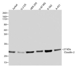
- Western blot analysis of Claudin-2 was performed by loading 20 µg of Jurkat, U-2 OS, HEK-293, U-87 MG, K562 and A431 cell lysates using Novex®NuPAGE®4-12% Bis-Tris gel (Product # NP0321BOX), XCell SureLock Electrophoresis System (Product # EI0002), Novex® Sharp Pre-Stained Protein Standard (Product # LC5800), and iBlot® Dry Blotting System (Product # IB21001). Proteins were transferred to a nitrocellulose membrane and blocked with 5% skim milk for 1 hour at room temperature. Claudin-2 was detected at ~27 kDa using Claudin-2 Recombinant Rabbit Polyclonal Antibody (Product # 710221) at a 1:1000 dilution in 2.5% skim milk at 4°C overnight on a rocking platform. Detection was performed using an HRP-conjugated Goat anti-Rabbit secondary antibody (Product # G-21234) at a 1:5000 dilution and chemiluminescent detection was performed using Pierce™ ECL Western blotting Substrate (Product # 32106).

- Submitted by
- Invitrogen Antibodies (provider)
- Main image

- Experimental details
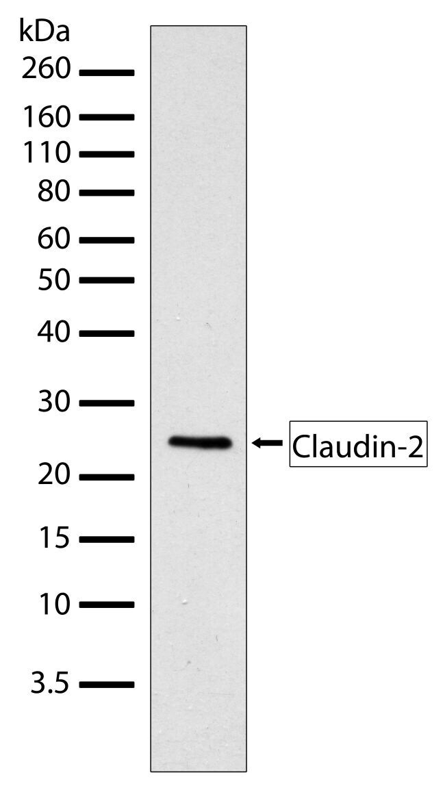
- Western blot analysis of Claudin-2 in whole cell extracts of MCF-7 using a Claudin-2 Recombinant Rabbit Polyclonal Antibody (Product # 710221) at a dilution of 2 µg/mL. Samples were detected using chemiluminescence (ECL). Results show a band at ~25kDa.
Supportive validation
- Submitted by
- Invitrogen Antibodies (provider)
- Main image

- Experimental details
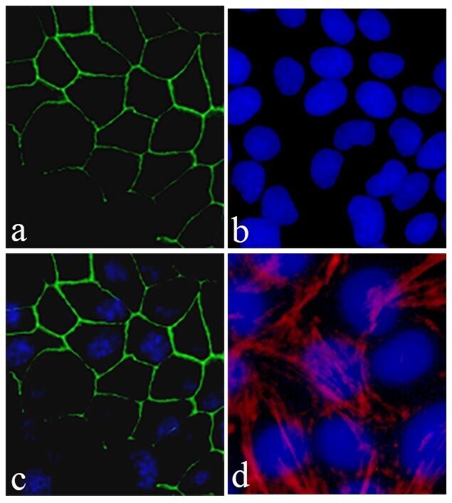
- Immunofluorescent analysis of Claudin-2 was performed on 90% confluent log phase Caco-2 cells. The cells were fixed with 4% paraformaldehyde for 15 minutes, and blocked with 5% BSA for 1 hour at room temperature. The cells were labeled with Claudin-2 Recombinant Rabbit Polyclonal Antibody (Product # 710221) at a dilution of 1:400 in 1% BSA and incubated for 3 hours at room temperature and then labeled with Alexa Fluor® 488 Goat anti-Rabbit IgG secondary antibody (Product # A-11008) at a dilution of 1:400 for 30 minutes at room temperature (Panel a: green). Nuclei (Panel b: blue) were stained with SlowFade® Gold Antifade Mountant with DAPI (Product # S36938). Panel c is a merged image showing cell junction localization and panel d is a control without primary antibody. The images were captured using a Nikon microscope at 20X magnification.
- Submitted by
- Invitrogen Antibodies (provider)
- Main image

- Experimental details
- Immunofluorescent analysis of Claudin-2 in HEK293 cells using a Claudin-2 Recombinant Rabbit Polyclonal Antibody (Product # 710221) followed by detection using an Alexa Fluor 488-conjugated Goat anti-Rabbit secondary antibody (green) (Image A). Nuclei were stained using DAPI (Image B) and actin stained with Alexa Fluor 594 phalloidin (red) (image C). Image D is a composite image showing localization of claudin-2 at the cell junction.
