Explore
 Validate
 Learn
Learn Western blot
Antibody data
- Antibody Data
- Antigen structure
- References [2]
- Comments [0]
- Validations
- Western blot [2]
- Immunohistochemistry [2]
- Other assay [2]
Submit
Validation data
Reference
Comment
Report error
- Product number
- PA1-37471 - Provider product page

- Provider
- Invitrogen Antibodies
- Product name
- Claudin 4 Polyclonal Antibody
- Antibody type
- Polyclonal
- Antigen
- Synthetic peptide
- Description
- This antibody is predicted to react with bovine, canine, amphibian, mouse, porcine and rat based on sequence homology.
- Concentration
- 2 mg/mL
Submitted references Investigating mammary glands of lactating goats for the presence of tertiary lymphoid organs.
MyD88 regulates a prolonged adaptation response to environmental dust exposure-induced lung disease.
Tsugami Y, Nakayama S, Suzuki N, Nii T, Isobe N
Frontiers in immunology 2022;13:941333
Frontiers in immunology 2022;13:941333
MyD88 regulates a prolonged adaptation response to environmental dust exposure-induced lung disease.
Johnson AN, Harkema JR, Nelson AJ, Dickinson JD, Kalil J, Duryee MJ, Thiele GM, Kumar B, Singh AB, Gaurav R, Glover SC, Tang Y, Romberger DJ, Kielian T, Poole JA
Respiratory research 2020 Apr 22;21(1):97
Respiratory research 2020 Apr 22;21(1):97
No comments: Submit comment
Supportive validation
- Submitted by
- Invitrogen Antibodies (provider)
- Main image
- Experimental details
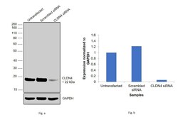
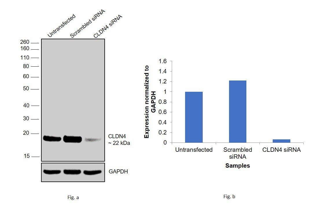
- Knockdown of CLDN7 was achieved by transfecting MCF7 with CLDN4 specific siRNAs (Silencer® select Product # s223316, s3441). Western blot analysis (Fig. a) was performed using membrane enriched extracts from knockdown cells (Lane 3), non-specific scrambled siRNA transfected cells (Lane 2) and untransfected cells (Lane 1). The blot was probed with Claudin 4 Polyclonal Antibody (Product # PA5-32354, 1:3000 dilution) and Goat anti-Rabbit IgG (H+L), Superclonal™ Recombinant Secondary Antibody, HRP (Product # A27036, 1:4000 dilution). Densitometric analysis of this western blot is shown in histogram (Fig. b). Decrease in signal upon siRNA mediated knock down confirms that antibody is specific to CLDN4.
- Submitted by
- Invitrogen Antibodies (provider)
- Main image
- Experimental details
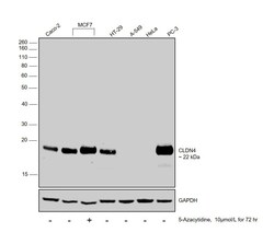
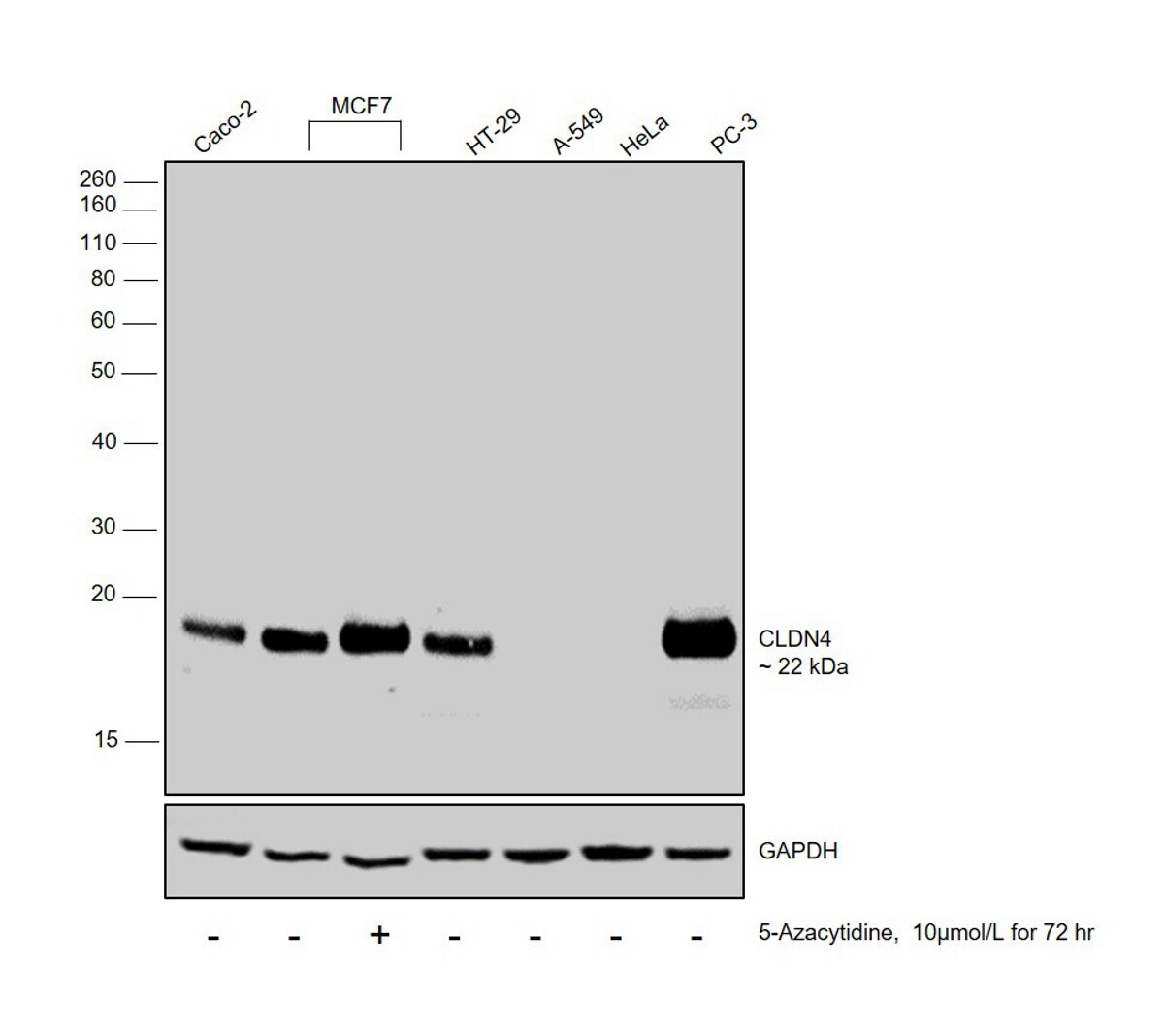
- Western blot was performed using Anti-Claudin 4 Polyclonal Antibody (Product # PA5-32354) and a 22 kDa band corresponding to CLDN4 was observed in Caco-2, MCF7, HT-29 and PC-3, not in A549 and HeLa which are reported to be negative for CLDN4 along with increased expression of the protein upon 5-Azacytidine treatment in MCF7. Membrane enriched extracts (30 µg lysate) of Caco-2 (Lane 1), MCF7 (Lane 2), MCF7 treated with 5-Azacytidine (Lane 3), HT-29 (Lane 4), A549 (Lane 5), HeLa (Lane 6) and PC-3 (Lane 7) were electrophoresed using Novex® NuPAGE® 12 % Bis-Tris gel (Product # NP0342BOX). Resolved proteins were then transferred onto a nitrocellulose membrane (Product # IB23001) by iBlot® 2 Dry Blotting System (Product # IB21001). The blot was probed with the primary antibody (1:1200 dilution) and detected by chemiluminescence with Goat anti-Rabbit IgG (H+L), Superclonal™ Recombinant Secondary Antibody, HRP (Product # A27036, 1:4000 dilution) using the iBright FL 1000 (Product # A32752). Chemiluminescent detection was performed using Novex® ECL Chemiluminescent Substrate Reagent Kit (Product # WP20005).
Supportive validation
- Submitted by
- Invitrogen Antibodies (provider)
- Main image
- Experimental details
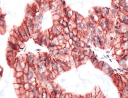
- Immunohistochemical analysis of Claudin-4 using anti-Claudin-4 Polyclonal Antibody (Product # PA5-32354) in Ovarian Carcinoma Cancer Tissue. The recommened dilution for this antibody in immunohistochemistry applications is 1:200.

- Submitted by
- Invitrogen Antibodies (provider)
- Main image
- Experimental details
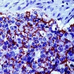
- Immunohistochemical analysis of Claudin 4 using a polyclonal antibody (Product # PA1-37471).

Supportive validation
- Submitted by
- Invitrogen Antibodies (provider)
- Main image

- Experimental details
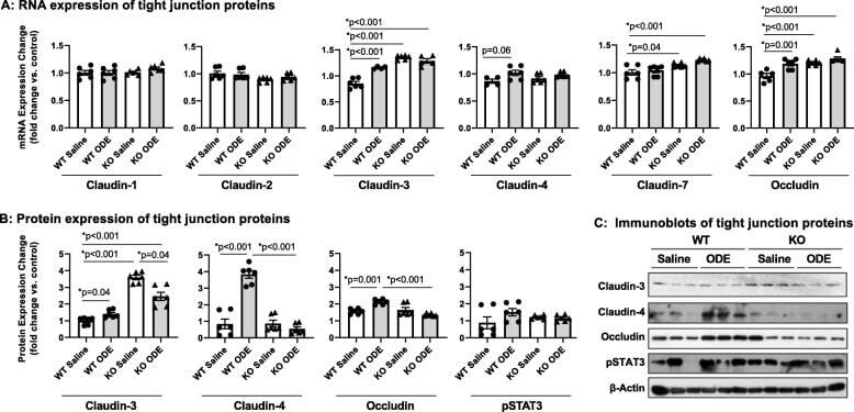
- Fig. 6 Repetitive ODE exposure increases expression of several tight junction proteins known to be upregulated in inflamed/injured lung in WT mice but not in MyD88 KO mice. WT and MyD88 KO mice were treated i.n. daily for 3 weeks with saline or ODE. Panel a, Expression of tight junction mRNA was measured by real-time quantitative PCR and are reported as fold-changes normalized to control. Panel b , Quantification of tight junction protein expression in ODE-treated mice compared to control mice as determined by the immunoblot presented in Panel c . Scatter plots demonstrate mean with standard error bars of N = 3 animals per group with 2 replicates per sample
- Submitted by
- Invitrogen Antibodies (provider)
- Main image

- Experimental details
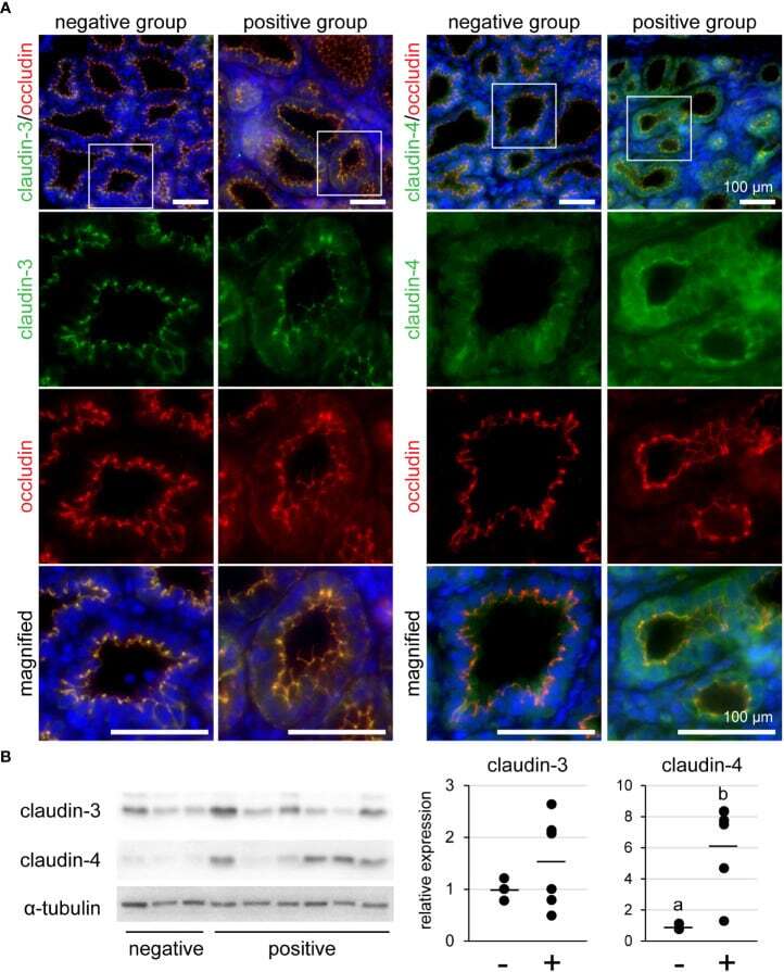
- Figure 5 (A) Representative images of immunofluorescence against claudin-3 and claudin-4 in tertiary lymphoid organ-positive and -negative goat mammary gland tissues. Occludin is a marker for tight junctions. Scale bar, 100 mum. (B) Bands detected by western blot and the results of densitometry analyses. Alpha-tubulin serves as internal control; (-) lymphocyte aggregation-negative group; (+) lymphocyte aggregation-positive group. Different letters indicate a significant difference between groups (p < 0.05).