Explore
 Validate
 Learn
Learn Western blot
 Immunocytochemistry
Antibody data
- Antibody Data
- Antigen structure
- References [3]
- Comments [0]
- Validations
- Immunocytochemistry [4]
- Other assay [3]
Submit
Validation data
Reference
Comment
Report error
- Product number
- PA5-20089 - Provider product page

- Provider
- Invitrogen Antibodies
- Product name
- Bim Polyclonal Antibody
- Antibody type
- Polyclonal
- Antigen
- Synthetic peptide
- Description
- A suggested positive control is K562 cell lysate. PA5-20089 can be used with blocking peptide PEP-0207. The PA5-20089 immunogen is located within amino acids 80-130 of BIM. Predicted molecular ~ 22kD. In Western blot applications, this antibody has been observed to detect a band at: 22kD. Predicted species reactivity based on immunogen sequence: Rat: (100%).
- Reactivity
- Human, Mouse
- Host
- Rabbit
- Isotype
- IgG
- Vial size
- 100 μg
- Concentration
- 1 mg/mL
- Storage
- Maintain refrigerated at 2-8°C for up to 3 months. For long term storage store at -20°C
Submitted references Oral Pathobiont Activates Anti-Apoptotic Pathway, Promoting both Immune Suppression and Oncogenic Cell Proliferation.
Human IDO-competent, long-lived immunoregulatory dendritic cells induced by intracellular pathogen, and their fate in humanized mice.
Nanostructured lipid carrier mediates effective delivery of methotrexate to induce apoptosis of rheumatoid arthritis via NF-κB and FOXO1.
Arjunan P, Meghil MM, Pi W, Xu J, Lang L, El-Awady A, Sullivan W, Rajendran M, Rabelo MS, Wang T, Tawfik OK, Kunde-Ramamoorthy G, Singh N, Muthusamy T, Susin C, Teng Y, Arce RM, Cutler CW
Scientific reports 2018 Nov 9;8(1):16607
Scientific reports 2018 Nov 9;8(1):16607
Human IDO-competent, long-lived immunoregulatory dendritic cells induced by intracellular pathogen, and their fate in humanized mice.
Tyagi RK, Miles B, Parmar R, Garg NK, Dalai SK, Baban B, Cutler CW
Scientific reports 2017 Feb 15;7:41083
Scientific reports 2017 Feb 15;7:41083
Nanostructured lipid carrier mediates effective delivery of methotrexate to induce apoptosis of rheumatoid arthritis via NF-κB and FOXO1.
Garg NK, Tyagi RK, Singh B, Sharma G, Nirbhavane P, Kushwah V, Jain S, Katare OP
International journal of pharmaceutics 2016 Feb 29;499(1-2):301-320
International journal of pharmaceutics 2016 Feb 29;499(1-2):301-320
No comments: Submit comment
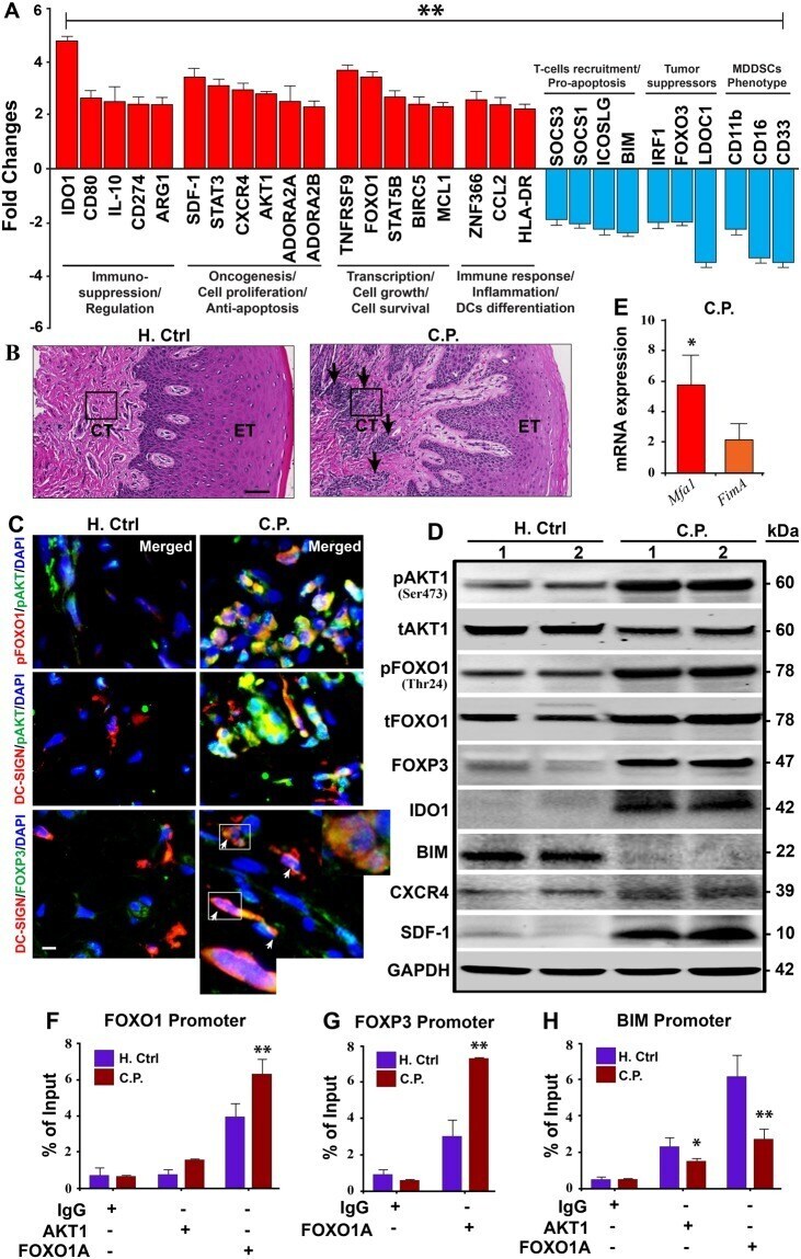
Supportive validation
- Submitted by
- Invitrogen Antibodies (provider)
- Main image
- Experimental details
- Immunocytochemistry of K562 cells using Bim Polyclonal Antibody (Product # PA5-20089) at 10 µg/mL. Cells were fixed with formaldehyde and blocked with 0.1 serum for 1 h at RT; antigen retrieval was by heat mediation with a citrate buffer (pH6). Samples were incubated with primary antibody overnight at 4°C. A goat anti-rabbit IgG H&L (HRP) at 1:250 was used as secondary. Counter stained with Hematoxylin.

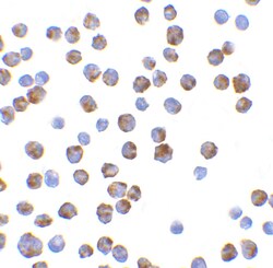
- Submitted by
- Invitrogen Antibodies (provider)
- Main image

- Experimental details
- Immunofluorescent analysis of 4% paraformaldehyde-fixed K562 cells labeling BIM with Bim Polyclonal Antibody (Product # PA5-20089) at 20 µg/mL, followed by goat anti-rabbit IgG secondary antibody at 1:500 dilution (red).

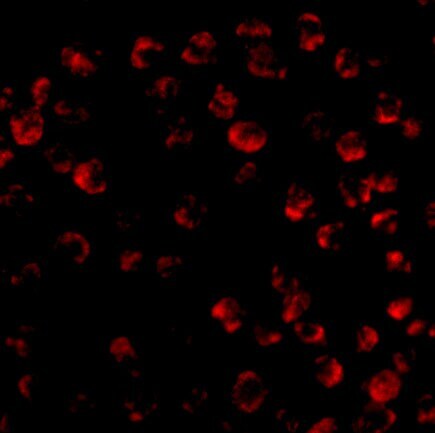
- Submitted by
- Invitrogen Antibodies (provider)
- Main image

- Experimental details
- Immunocytochemistry of K562 cells using Bim Polyclonal Antibody (Product # PA5-20089) at 10 µg/mL. Cells were fixed with formaldehyde and blocked with 0.1 serum for 1 h at RT; antigen retrieval was by heat mediation with a citrate buffer (pH6). Samples were incubated with primary antibody overnight at 4°C. A goat anti-rabbit IgG H&L (HRP) at 1:250 was used as secondary. Counter stained with Hematoxylin.
Supportive validation
- Submitted by
- Invitrogen Antibodies (provider)
- Main image
- Experimental details
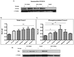
- Figure 2 Phosphorylated Foxo1 decreases with PDDC apoptosis rate. ( A ) FOXO1 expression was observed on protein level in PDDCs (WT & DPG) by carrying out Western blot using Anti-FOXO1 Mab. alpha-Tubulin, loading control (n = 2). The amount of total PDDC Foxo1 ( B ) and phosphorylated Foxo1 ( C ) were measured by whole cell ELISA and normalized to total cell numbers (n = 3) (B) PDDCs generated with E. coli LPS or either of the P. gingivalis strains have significantly higher levels of Foxo1. ( C ) PDDCs generated by the minor-fimbriae expressing WT and DPG-3 or the non-fimbriated MFB have significantly higher levels of phosphorylated Foxo1 ( D ) DCs were washed in RPMI and then incubated for 10 hrs in 10% FBS in RPMI. DCs were then lysed and Bim was detected by immunoblot. alpha-tubulin, loading control (n = 2).

- Submitted by
- Invitrogen Antibodies (provider)
- Main image
- Experimental details
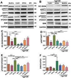
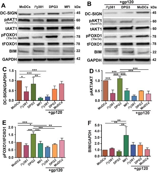
- Figure 1 DPG3 activated DC-SIGN, AKT, regulated FOXO1 phosphorylation/expression was required for the survival effect of DPG3 in MDDSCs. Immature monocytes isolated from human peripheral blood mononuclear cells (PBMCs) were infected with Pg 381, DPG3 and MFI for 6 hours at 1 MOI. ( A ) Immunoblot analysis of DC-SIGN, p/tAKT, p/tFOXO1 and BIM in MDDSCs and MoDCs with GAPDH as loading control. Immunoblot detected increased expression of DC-SIGN, p/tAKT, p/tFOXO1 and decreased expression of BIM in DPG3- infected MDDSCs compared with uninfected control MoDCs. ( B ) DC-SIGN was blocked with HIV-gp120 (6ug/ml) for 30 minutes before infection. Notably, DC-SIGN expression and internalization was abolished by its antagonist HIV-gp120 and consequently the downstream signaling cascade also blocked as evident by dephosphorylated or decreased expression of pAKT ( B , D ), pFOXO1 ( B , E ) and increased expression of BIM ( B , F ). ( C - F ) Quantification analysis of DC-SIGN ( C ), and relative ratio of p/tAKT ( D ), p/tFOXO1 ( E ) and BIM ( F ) after normalized with the internal control GAPDH. Results are from one representative of three independent experiments. Data were analyzed by performing one-way ANOVA with Tukey's post hoc using Prism GraphPad. * P
- Submitted by
- Invitrogen Antibodies (provider)
- Main image
- Experimental details
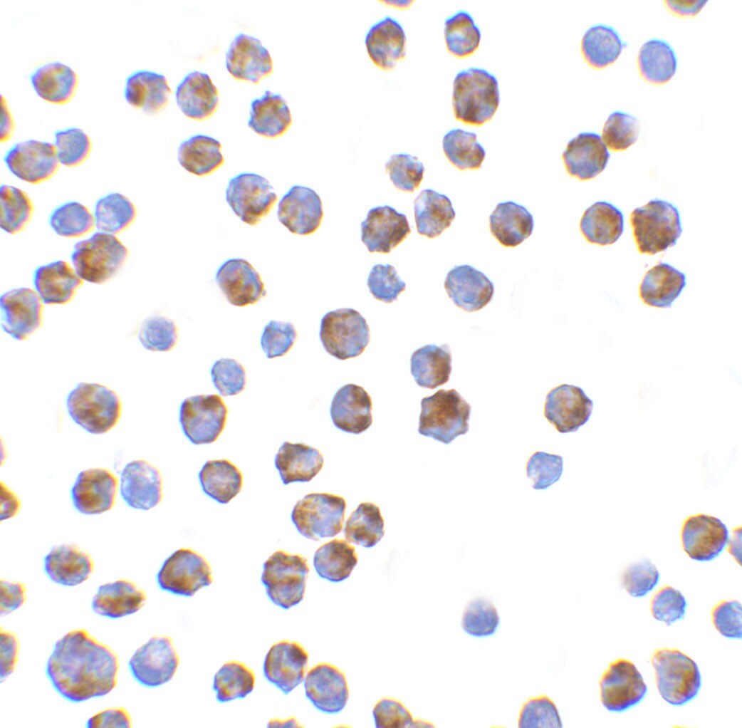
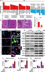
- Figure 4 Enriched blood panDCs and gingival tissues of CP patients reveal immunosuppressive state. ( A ) 29 genes associated with transcriptional regulation, inflammation, immunosuppression, cell proliferation, anti-apoptosis and other homeostatic functions were identified from the RNAseq data. Bar graphs show both up- (red) and down- (blue) regulation of genes (>=+-2 fold) in ex-vivo isolated and panDC-enriched blood samples from CP patients compared to healthy controls confirmed by TaqManPCR. The overall validation data shows the same trend as in RNAseq analysis. ( B ) H&E of healthy control and CP patient's gingival tissue. The arrows indicate the infiltrating cells of inflammatory responses; (20X), CT- Connective Tissue, ET- Epithelial Tissue. Results are representative of two experiments (n = 9/group). ( C ) Co-localized expression of pAKT1/pFOXO1, pAKT1/DC-SIGN (receptor for Mfa1) and DC-SIGN/FOXP3 (arrowhead) in gingival connective tissue (marked in b) from CP, compared with healthy control. Images are representative of three independent experiments (Scale bar- 100 um; pls refer Fig. S6 for independent channel and their quantification measurement of co-localization. ( D ) Immunoblot analysis of p/tAKT1, p/tFOXO1, FOXP3, IDO1, BIM, CXCR4 and SDF1 in gingival tissue of CP compared with healthy control. ( E ) Mfa1 and FimA mRNA in ex-vivo isolated panDCs from CP patients and Healthy control and normalized with the internal control. ( F-H ) Regulation of FOXO1, FOXP3, BIM