Explore
 Validate
 Learn
Learn Western blot
Antibody data
- Antibody Data
- Antigen structure
- References [3]
- Comments [0]
- Validations
- Western blot [1]
- Immunocytochemistry [1]
- Immunohistochemistry [1]
Submit
Validation data
Reference
Comment
Report error
- Product number
- AM1829b - Provider product page

- Provider
- Abcepta
- Proper citation
- Abgent Cat#AM1829b, RRID:AB_10664137
- Product name
- Beta-Actin Antibody
- Antibody type
- Monoclonal
- Antigen
- Recombinant Protein
- Description
- Mouse Monoclonal Antibody (Mab)
- Reactivity
- Human, Mouse
- Host
- Mouse
- Isotype
- IgG
- Vial size
- 400 µl
- Concentration
- 0.5 mg/ml
- Storage
- Maintain refrigerated at 2-8°C for up to 6 months. For long term storage store at -20°C in small aliquots to prevent freeze-thaw cycles.
Submitted references Evidence for a novel antioxidant function and isoform-specific regulation of the human p66Shc gene.
Functional analyses of the three simian hemorrhagic fever virus nonstructural protein 1 papain-like proteases.
Addiction to multiple oncogenes can be exploited to prevent the emergence of therapeutic resistance.
Miyazawa M, Tsuji Y
Molecular biology of the cell 2014 Jul 1;25(13):2116-27
Molecular biology of the cell 2014 Jul 1;25(13):2116-27
Functional analyses of the three simian hemorrhagic fever virus nonstructural protein 1 papain-like proteases.
Vatter HA, Di H, Donaldson EF, Radu GU, Maines TR, Brinton MA
Journal of virology 2014 Aug;88(16):9129-40
Journal of virology 2014 Aug;88(16):9129-40
Addiction to multiple oncogenes can be exploited to prevent the emergence of therapeutic resistance.
Choi PS, Li Y, Felsher DW
Proceedings of the National Academy of Sciences of the United States of America 2014 Aug 12;111(32):E3316-24
Proceedings of the National Academy of Sciences of the United States of America 2014 Aug 12;111(32):E3316-24
No comments: Submit comment
Supportive validation
- Submitted by
- Abcepta (provider)
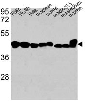
- Main image

- Experimental details
- "Western blot analysis of anti-ACTB Antibody (Cat. #AM1829b) in K562, HL-60,Hela cell line, mouse spleen, mouse liver tissue lysates, mouse NIH-3T3 cell line lysate and mouse cerebellum, mouse brain tissue lysates (35ug/lane). ACTB (arrow) was detected using the purified Mab. "
- Primary Ab dilution
- 1:1000
Supportive validation
- Submitted by
- Abcepta (provider)
- Main image

- Experimental details
- Confocal immunofluorescent analysis of ACTB Antibody (Cat#AM1829b) with Hela cell followed by Alexa Fluor 488-conjugated goat anti-mouse lgG (green). DAPI was used to stain the cell nuclear (blue).
- Primary Ab dilution
- 1:10~50

Supportive validation
- Submitted by
- Abcepta (provider)
- Main image
- Experimental details
- ACTB Antibody (AM1829b)immunohistochemistry analysis in formalin fixed and paraffin embedded human skeletal muscle followed by peroxidase conjugation of the secondary antibody and DAB staining.This data demonstrates the use of ACTB Antibody for immunohistochemistry. Clinical relevance has not been evaluated.
- Primary Ab dilution
- 1:10~50