Explore
 Validate
 Learn
Learn Western blot
 Immunocytochemistry
Antibody data
- Antibody Data
- Antigen structure
- References [30]
- Comments [0]
- Validations
- Western blot [26]
- Immunocytochemistry [2]
- Immunoprecipitation [1]
- Immunohistochemistry [3]
Submit
Validation data
Reference
Comment
Report error
- Product number
- GTX110564 - Provider product page

- Provider
- GeneTex
- Proper citation
- GeneTex Cat#GTX110564, RRID:AB_10618080
- Product name
- beta Actin antibody
- Antibody type
- Polyclonal
- Reactivity
- Human, Mouse, Rat, Drosophila
- Host
- Rabbit
Submitted references Deletion of long noncoding RNA EFNA3 aggravates hypoxia-induced injury in PC-12 cells by upregulation of miR-101a.
Characterizing the protective effects of SHLP2, a mitochondrial-derived peptide, in macular degeneration.
Homoharringtonine induced immune alteration for an Efficient Anti-tumor Response in Mouse Models of Non-small Cell Lung Adenocarcinoma Expressing Kras Mutation.
Temporal Expression Patterns of Clock Genes and Aquaporin 5/Anoctamin 1 in Rat Submandibular Gland Cells.
CCL5 is induced by TLR 3 signaling in HuCCT1 human biliary epithelial cells: possible involvement in the pathogenesis of biliary atresia.
Interferon (IFN)-induced protein 35 (IFI35) negatively regulates IFN-β-phosphorylated STAT1-RIG-I-CXCL10/CCL5 axis in U373MG astrocytoma cells treated with polyinosinic-polycytidylic acid.
CYT-Rx20 inhibits ovarian cancer cells in vitro and in vivo through oxidative stress-induced DNA damage and cell apoptosis.
Synthetic β-nitrostyrene derivative CYT-Rx20 as inhibitor of oral cancer cell proliferation and tumor growth through glutathione suppression and reactive oxygen species induction.
A comparative study of the influence of two types of PHEMA stents on the differentiation of ASCs to myocardial cells.
Endogenous reactive oxygen species cause astrocyte defects and neuronal dysfunctions in the hippocampus: a new model for aging brain.
Functional analysis of a novel, thyroglobulin-embedded microRNA gene deregulated in papillary thyroid carcinoma.
Differential Expression of Complement Markers in Normal and AMD Transmitochondrial Cybrids.
Family of microRNA-146 Regulates RARβ in Papillary Thyroid Carcinoma.
Inhibition of DNA methyltransferase 1 increases nuclear receptor subfamily 4 group A member 1 expression and decreases blood glucose in type 2 diabetes.
Down-regulated and Commonly mutated ALPK1 in Lung and Colorectal Cancers.
Discovery of BPR1K871, a quinazoline based, multi-kinase inhibitor for the treatment of AML and solid tumors: Rational design, synthesis, in vitro and in vivo evaluation.
KLHL39 suppresses colon cancer metastasis by blocking KLHL20-mediated PML and DAPK ubiquitination.
PTPRD silencing by DNA hypermethylation decreases insulin receptor signaling and leads to type 2 diabetes.
The Differential Profiling of Ubiquitin-Proteasome and Autophagy Systems in Different Tissues before the Onset of Huntington's Disease Models.
Dimethyl sulfoxide inhibits spontaneous diabetes and autoimmune recurrence in non-obese diabetic mice by inducing differentiation of regulatory T cells.
Targeting the human androgen receptor gene with platinated triplex-forming oligonucleotides.
Enhanced antitumor efficacy of an oncolytic herpes simplex virus expressing an endostatin-angiostatin fusion gene in human glioblastoma stem cell xenografts.
Evaluation of the antitumor effects of BPR1J-340, a potent and selective FLT3 inhibitor, alone or in combination with an HDAC inhibitor, vorinostat, in AML cancer.
Genetically induced oxidative stress in mice causes thrombocytosis, splenomegaly and placental angiodysplasia that leads to recurrent abortion.
Radiation sensitization of tumor cells induced by shear stress: the roles of integrins and FAK.
The active zone protein family ELKS supports Ca2+ influx at nerve terminals of inhibitory hippocampal neurons.
GALNT2 enhances migration and invasion of oral squamous cell carcinoma by regulating EGFR glycosylation and activity.
Autophagy promotes resistance to photodynamic therapy-induced apoptosis selectively in colorectal cancer stem-like cells.
Antiosteoclastogenic activity of silicate-based materials antagonizing receptor activator for nuclear factor kappaB ligand-induced osteoclast differentiation of murine marcophages.
Amyloid-beta (Aβ) D7H mutation increases oligomeric Aβ42 and alters properties of Aβ-zinc/copper assemblies.
Gong W, Qie S, Huang P, Xi J
Journal of cellular biochemistry 2019 Jan;120(1):836-847
Journal of cellular biochemistry 2019 Jan;120(1):836-847
Characterizing the protective effects of SHLP2, a mitochondrial-derived peptide, in macular degeneration.
Nashine S, Cohen P, Nesburn AB, Kuppermann BD, Kenney MC
Scientific reports 2018 Oct 11;8(1):15175
Scientific reports 2018 Oct 11;8(1):15175
Homoharringtonine induced immune alteration for an Efficient Anti-tumor Response in Mouse Models of Non-small Cell Lung Adenocarcinoma Expressing Kras Mutation.
Weng TY, Wu HF, Li CY, Hung YH, Chang YW, Chen YL, Hsu HP, Chen YH, Wang CY, Chang JY, Lai MD
Scientific reports 2018 May 29;8(1):8216
Scientific reports 2018 May 29;8(1):8216
Temporal Expression Patterns of Clock Genes and Aquaporin 5/Anoctamin 1 in Rat Submandibular Gland Cells.
Satou R, Sato M, Kimura M, Ishizuka Y, Tazaki M, Sugihara N, Shibukawa Y
Frontiers in physiology 2017;8:320
Frontiers in physiology 2017;8:320
CCL5 is induced by TLR 3 signaling in HuCCT1 human biliary epithelial cells: possible involvement in the pathogenesis of biliary atresia.
Shimada T, Imaizumi T, Shirai K, Tatsuta T, Kimura T, Hayakari R, Yoshida H, Matsumiya T, Kijima H, Mizukami H, Hakamada K
Biomedical research (Tokyo, Japan) 2017;38(5):269-276
Biomedical research (Tokyo, Japan) 2017;38(5):269-276
Interferon (IFN)-induced protein 35 (IFI35) negatively regulates IFN-β-phosphorylated STAT1-RIG-I-CXCL10/CCL5 axis in U373MG astrocytoma cells treated with polyinosinic-polycytidylic acid.
Shirai K, Shimada T, Yoshida H, Hayakari R, Matsumiya T, Tanji K, Murakami M, Tanaka H, Imaizumi T
Brain research 2017 Mar 1;1658:60-67
Brain research 2017 Mar 1;1658:60-67
CYT-Rx20 inhibits ovarian cancer cells in vitro and in vivo through oxidative stress-induced DNA damage and cell apoptosis.
Wang YY, Chen YK, Hu SC, Hsu YL, Tsai CH, Chi TC, Huang WL, Hsieh PW, Yuan SF
Cancer chemotherapy and pharmacology 2017 Jun;79(6):1129-1140
Cancer chemotherapy and pharmacology 2017 Jun;79(6):1129-1140
Synthetic β-nitrostyrene derivative CYT-Rx20 as inhibitor of oral cancer cell proliferation and tumor growth through glutathione suppression and reactive oxygen species induction.
Wang YY, Chen YK, Hsu YL, Chiu WC, Tsai CH, Hu SC, Hsieh PW, Yuan SF
Head & neck 2017 Jun;39(6):1055-1064
Head & neck 2017 Jun;39(6):1055-1064
A comparative study of the influence of two types of PHEMA stents on the differentiation of ASCs to myocardial cells.
Lao S, Xu J, Liu Y, Cai S, Lin L, Zhang J, Cai D, Yin S
Molecular medicine reports 2017 Jul;16(1):507-514
Molecular medicine reports 2017 Jul;16(1):507-514
Endogenous reactive oxygen species cause astrocyte defects and neuronal dysfunctions in the hippocampus: a new model for aging brain.
Ishii T, Takanashi Y, Sugita K, Miyazawa M, Yanagihara R, Yasuda K, Onouchi H, Kawabe N, Nakata M, Yamamoto Y, Hartman PS, Ishii N
Aging cell 2017 Feb;16(1):39-51
Aging cell 2017 Feb;16(1):39-51
Functional analysis of a novel, thyroglobulin-embedded microRNA gene deregulated in papillary thyroid carcinoma.
Kolanowska M, Wójcicka A, Kubiak A, Świerniak M, Kotlarek M, Maciąg M, Gaj P, Koperski Ł, Górnicka B, Jażdżewski K
Scientific reports 2017 Aug 30;7(1):9942
Scientific reports 2017 Aug 30;7(1):9942
Differential Expression of Complement Markers in Normal and AMD Transmitochondrial Cybrids.
Nashine S, Chwa M, Kazemian M, Thaker K, Lu S, Nesburn A, Kuppermann BD, Kenney MC
PloS one 2016;11(8):e0159828
PloS one 2016;11(8):e0159828
Family of microRNA-146 Regulates RARβ in Papillary Thyroid Carcinoma.
Czajka AA, Wójcicka A, Kubiak A, Kotlarek M, Bakuła-Zalewska E, Koperski Ł, Wiechno W, Jażdżewski K
PloS one 2016;11(3):e0151968
PloS one 2016;11(3):e0151968
Inhibition of DNA methyltransferase 1 increases nuclear receptor subfamily 4 group A member 1 expression and decreases blood glucose in type 2 diabetes.
Chen YT, Liao JW, Tsai YC, Tsai FJ
Oncotarget 2016 Jun 28;7(26):39162-39170
Oncotarget 2016 Jun 28;7(26):39162-39170
Down-regulated and Commonly mutated ALPK1 in Lung and Colorectal Cancers.
Liao HF, Lee HH, Chang YS, Lin CL, Liu TY, Chen YC, Yen JC, Lee YT, Lin CY, Wu SH, Ko YC, Chang JG
Scientific reports 2016 Jun 10;6:27350
Scientific reports 2016 Jun 10;6:27350
Discovery of BPR1K871, a quinazoline based, multi-kinase inhibitor for the treatment of AML and solid tumors: Rational design, synthesis, in vitro and in vivo evaluation.
Hsu YC, Coumar MS, Wang WC, Shiao HY, Ke YY, Lin WH, Kuo CC, Chang CW, Kuo FM, Chen PY, Wang SY, Li AS, Chen CH, Kuo PC, Chen CP, Wu MH, Huang CL, Yen KJ, Chang YI, Hsu JT, Chen CT, Yeh TK, Song JS, Shih C, Hsieh HP
Oncotarget 2016 Dec 27;7(52):86239-86256
Oncotarget 2016 Dec 27;7(52):86239-86256
KLHL39 suppresses colon cancer metastasis by blocking KLHL20-mediated PML and DAPK ubiquitination.
Chen HY, Hu JY, Chen TH, Lin YC, Liu X, Lin MY, Lang YD, Yen Y, Chen RH
Oncogene 2015 Oct 1;34(40):5141-51
Oncogene 2015 Oct 1;34(40):5141-51
PTPRD silencing by DNA hypermethylation decreases insulin receptor signaling and leads to type 2 diabetes.
Chen YT, Lin WD, Liao WL, Lin YJ, Chang JG, Tsai FJ
Oncotarget 2015 May 30;6(15):12997-3005
Oncotarget 2015 May 30;6(15):12997-3005
The Differential Profiling of Ubiquitin-Proteasome and Autophagy Systems in Different Tissues before the Onset of Huntington's Disease Models.
Her LS, Lin JY, Fu MH, Chang YF, Li CL, Tang TY, Jhang YL, Chang CY, Shih MC, Cheng PH, Yang SH
Brain pathology (Zurich, Switzerland) 2015 Jul;25(4):481-90
Brain pathology (Zurich, Switzerland) 2015 Jul;25(4):481-90
Dimethyl sulfoxide inhibits spontaneous diabetes and autoimmune recurrence in non-obese diabetic mice by inducing differentiation of regulatory T cells.
Lin GJ, Sytwu HK, Yu JC, Chen YW, Kuo YL, Yu CC, Chang HM, Chan DC, Huang SH
Toxicology and applied pharmacology 2015 Jan 15;282(2):207-14
Toxicology and applied pharmacology 2015 Jan 15;282(2):207-14
Targeting the human androgen receptor gene with platinated triplex-forming oligonucleotides.
Graham MK, Brown TR, Miller PS
Biochemistry 2015 Apr 7;54(13):2270-82
Biochemistry 2015 Apr 7;54(13):2270-82
Enhanced antitumor efficacy of an oncolytic herpes simplex virus expressing an endostatin-angiostatin fusion gene in human glioblastoma stem cell xenografts.
Zhang G, Jin G, Nie X, Mi R, Zhu G, Jia W, Liu F
PloS one 2014;9(4):e95872
PloS one 2014;9(4):e95872
Evaluation of the antitumor effects of BPR1J-340, a potent and selective FLT3 inhibitor, alone or in combination with an HDAC inhibitor, vorinostat, in AML cancer.
Lin WH, Yeh TK, Jiaang WT, Yen KJ, Chen CH, Huang CT, Yen SC, Hsieh SY, Chou LH, Chen CP, Chiu CH, Kao LC, Chao YS, Chen CT, Hsu JT
PloS one 2014;9(1):e83160
PloS one 2014;9(1):e83160
Genetically induced oxidative stress in mice causes thrombocytosis, splenomegaly and placental angiodysplasia that leads to recurrent abortion.
Ishii T, Miyazawa M, Takanashi Y, Tanigawa M, Yasuda K, Onouchi H, Kawabe N, Mitsushita J, Hartman PS, Ishii N
Redox biology 2014;2:679-85
Redox biology 2014;2:679-85
Radiation sensitization of tumor cells induced by shear stress: the roles of integrins and FAK.
Luo CW, Wu CC, Ch'ang HJ
Biochimica et biophysica acta 2014 Sep;1843(9):2129-37
Biochimica et biophysica acta 2014 Sep;1843(9):2129-37
The active zone protein family ELKS supports Ca2+ influx at nerve terminals of inhibitory hippocampal neurons.
Liu C, Bickford LS, Held RG, Nyitrai H, Südhof TC, Kaeser PS
The Journal of neuroscience : the official journal of the Society for Neuroscience 2014 Sep 10;34(37):12289-303
The Journal of neuroscience : the official journal of the Society for Neuroscience 2014 Sep 10;34(37):12289-303
GALNT2 enhances migration and invasion of oral squamous cell carcinoma by regulating EGFR glycosylation and activity.
Lin MC, Huang MJ, Liu CH, Yang TL, Huang MC
Oral oncology 2014 May;50(5):478-84
Oral oncology 2014 May;50(5):478-84
Autophagy promotes resistance to photodynamic therapy-induced apoptosis selectively in colorectal cancer stem-like cells.
Wei MF, Chen MW, Chen KC, Lou PJ, Lin SY, Hung SC, Hsiao M, Yao CJ, Shieh MJ
Autophagy 2014 Jul;10(7):1179-92
Autophagy 2014 Jul;10(7):1179-92
Antiosteoclastogenic activity of silicate-based materials antagonizing receptor activator for nuclear factor kappaB ligand-induced osteoclast differentiation of murine marcophages.
Hung CJ, Kao CT, Chen YJ, Shie MY, Huang TH
Journal of endodontics 2013 Dec;39(12):1557-61
Journal of endodontics 2013 Dec;39(12):1557-61
Amyloid-beta (Aβ) D7H mutation increases oligomeric Aβ42 and alters properties of Aβ-zinc/copper assemblies.
Chen WT, Hong CJ, Lin YT, Chang WH, Huang HT, Liao JY, Chang YJ, Hsieh YF, Cheng CY, Liu HC, Chen YR, Cheng IH
PloS one 2012;7(4):e35807
PloS one 2012;7(4):e35807
No comments: Submit comment
Enhanced validation
Supportive validation
- Submitted by
- GeneTex (provider)
- Enhanced method
- Genetic validation
- Main image
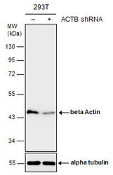
- Experimental details
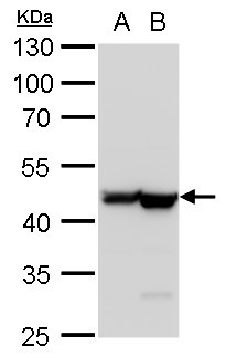
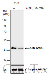
- Non-transfected (¡V) and transfected (+) 293T whole cell extracts (10 ?g) were separated by 10% SDS-PAGE, and the membrane was blotted with beta Actin antibody (GTX110564) diluted at 1:15000. The HRP-conjugated anti-rabbit IgG antibody (GTX213110-01) was used to detect the primary antibody.
Supportive validation
- Submitted by
- GeneTex (provider)
- Main image
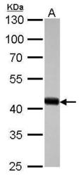
- Experimental details
- beta Actin antibody detects ACTB protein by western blot analysis.A. 30 ?g drosophila lysate/extract10% SDS-PAGEbeta Actin antibody (GTX110564) dilution: 1:10000 The HRP-conjugated anti-rabbit IgG antibody (GTX213110-01) was used to detect the primary antibody.

- Submitted by
- GeneTex (provider)
- Main image
- Experimental details
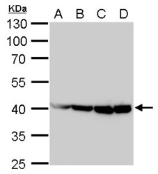
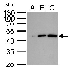
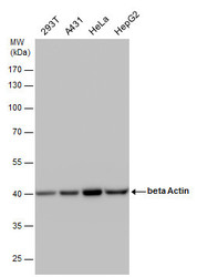
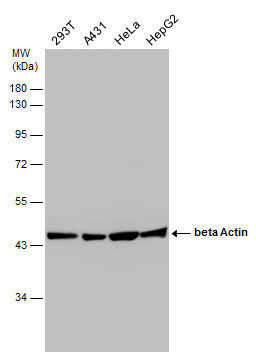
- beta Actin antibody detects beta Actin protein by Western blot analysis.A. 30 µg 293T whole cell lysate/extractB. 30 µg A431 whole cell lysate/extractC. 30 µg HeLa whole cell lysate/extractD. 30 µg HepG2 whole cell lysate/extract10 % S
- Validation comment
- WB

- Submitted by
- GeneTex (provider)
- Main image

- Experimental details
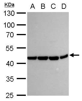
- beta Actin antibody detects beta Actin protein by Western blot analysis.A. 30 µg Neuro2A whole cell lysate/extractB. 30 µg GL261 whole cell lysate/extractC. 30 µg C8D30 whole cell lysate/extractD. 30 µg NIH-3T3 whole cell lysate/extract
- Validation comment
- WB
- Submitted by
- GeneTex (provider)
- Main image
- Experimental details
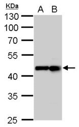
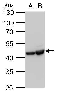
- beta Actin antibody detects beta Actin protein by Western blot analysis.A. 30 µg PC-12 whole cell lysate/extractB. 30 µg Rat-2 whole cell lysate/extract10 % SDS-PAGEbeta Actin antibody (GTX110564) dilution: 1:20000
- Validation comment
- WB

- Submitted by
- GeneTex (provider)
- Main image

- Experimental details
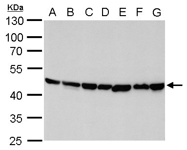
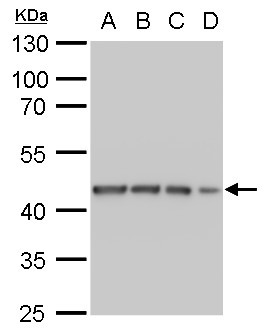
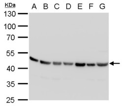
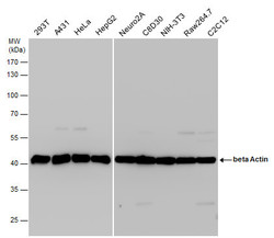
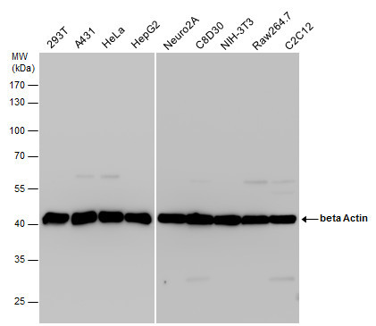
- beta Actin antibody detects beta Actin protein by western blot analysis.A. 30 ?g Neuro 2A whole cell lysate/extractB. 30 ?g GL261 whole cell lysate/extract C. 30 ?g C8D30 whole cell lysate/extractD. 30 ?g NIH-3T3 whole cell lysate/extractE. 30 ?g BCL-1 whole cell lysate/extract F. 30 ?g Raw264.7 whole cell lysate/extract G. 30 ?g C2C12 whole cell lysate/extract10 % SDS-PAGEbeta Actin antibody (GTX110564) dilution: 1:20000
- Validation comment
- WB
- Submitted by
- GeneTex (provider)
- Main image

- Experimental details
- beta Actin antibody detects beta Actin protein by western blot analysis.A. 30 ?g PC-12 whole cell lysate/extractB. 30 ?g Rat2 whole cell lysate/extract10 % SDS-PAGEbeta Actin antibody (GTX110564) dilution: 1:20000
- Validation comment
- WB
- Submitted by
- GeneTex (provider)
- Main image

- Experimental details
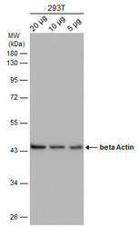
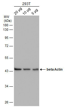
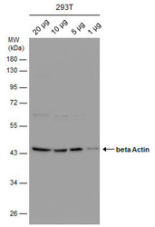
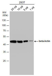
- beta Actin antibody detects beta Actin protein by western blot analysis.A. 1 ?g 293T whole cell lysate/extract B. 5 ?g 293T whole cell lysate/extract C. 10 ?g 293T whole cell lysate/extract10 % SDS-PAGEbeta Actin antibody (GTX110564) dilution: 1:20000
- Validation comment
- WB
- Submitted by
- GeneTex (provider)
- Main image

- Experimental details
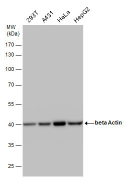
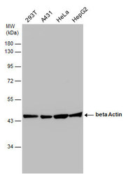
- beta Actin antibody detects beta Actin protein by western blot analysis.A. 30 ?g 293T whole cell lysate/extractB. 30 ?g A431 whole cell lysate/extract C. 30 ?g HeLa whole cell lysate/extractD. 30 ?g HepG2 whole cell lysate/extract10 % SDS-PAGEbeta Actin antibody (GTX110564) dilution: 1:20000
- Validation comment
- WB
- Submitted by
- GeneTex (provider)
- Main image

- Experimental details
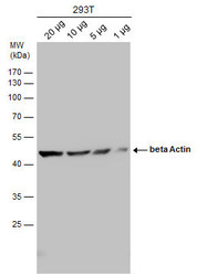
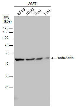
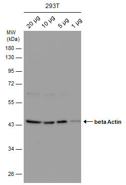
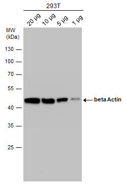
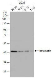
- beta Actin antibody detects beta Actin protein by western blot analysis.A. 20 ?g 293T whole cell lysate/extract B. 10 ?g 293T whole cell lysate/extract C. 5 ?g 293T whole cell lysate/extract D. 1 ?g 293T whole cell lysate/extract10 % SDS-PAGEbeta Actin antibody (GTX110564) dilution: 1:20000
- Validation comment
- WB
- Submitted by
- GeneTex (provider)
- Main image

- Experimental details
- beta Actin antibody detects beta Actin protein by western blot analysis.A. 30 ?g PC-12 whole cell lysate/extract B. 30 ?g Rat2 whole cell lysate/extract10% SDS-PAGEbeta Actin antibody (GTX110564) dilution: 1:20000The HRP-conjugated anti-rabbit IgG antibody (GTX213110-01) was used to detect the primary antibody.
- Submitted by
- GeneTex (provider)
- Main image
- Experimental details
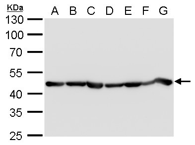
- beta Actin antibody detects beta Actin protein by western blot analysis.A. 30 ?g Neuro2A whole cell lysate/extract B. 30 ?g GL261 whole cell lysate/extract C. 30 ?g C8D30 whole cell lysate/extract D. 30 ?g NIH-3T3 whole cell lysate/extract E. 30 ?g BCL-1 whole cell lysate/extract F. 30 ?g Raw 264.7 whole cell lysate/extract G. 30 ?g C2Cl2 whole cell lysate/extract10 % SDS-PAGEbeta Actin antibody (GTX110564) dilution: 1:20000
- Validation comment
- WB

- Submitted by
- GeneTex (provider)
- Main image
- Experimental details
- beta Actin antibody detects beta Actin protein by western blot analysis. Various whole cell extracts (30 ?g) were separated by 10% SDS-PAGE, and the membrane was blotted with beta Actin antibody (GTX110564) diluted by 1:20000.
- Validation comment
- WB
- Submitted by
- GeneTex (provider)
- Main image
- Experimental details
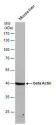
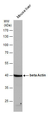
- beta Actin antibody detects beta Actin protein by western blot analysis. Mouse tissue extracts (50 ?g) was separated by 10% SDS-PAGE, and the membrane was blotted with beta Actin antibody (GTX110564) diluted by 1:20000. The HRP-conjugated anti-rabbit IgG antibody (GTX213110-01) was used to detect the primary antibody.
- Submitted by
- GeneTex (provider)
- Main image
- Experimental details
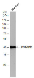
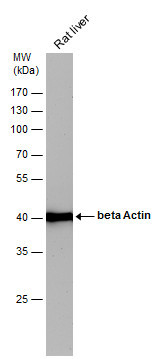
- beta Actin antibody detects beta Actin protein by western blot analysis. Rat tissue extracts (50 ?g) was separated by 10% SDS-PAGE, and the membrane was blotted with beta Actin antibody (GTX110564) diluted by 1:20000. The HRP-conjugated anti-rabbit IgG antibody (GTX213110-01) was used to detect the primary antibody.
- Submitted by
- GeneTex (provider)
- Main image
- Experimental details
- beta Actin antibody detects beta Actin protein by western blot analysis. Various whole cell extracts (30 ?g) were separated by 10% SDS-PAGE, and the membrane was blotted with beta Actin antibody (GTX110564) diluted at a dilution of 1:20000. The HRP-conjugated anti-rabbit IgG antibody (GTX213110-01) was used to detect the primary antibody.
- Submitted by
- GeneTex (provider)
- Main image
- Experimental details
- Various whole cell extracts (30 ?g) were separated by 10% SDS-PAGE, and the membrane was blotted with beta Actin antibody (GTX110564) diluted at 1:20000. The HRP-conjugated anti-rabbit IgG antibody (GTX213110-01) was used to detect the primary antibody.
- Submitted by
- GeneTex (provider)
- Main image
- Experimental details
- Various whole cell extracts were separated by 10% SDS-PAGE, and the membrane was blotted with beta Actin antibody (GTX110564) diluted at 1:20000.
- Submitted by
- GeneTex (provider)
- Main image
- Experimental details
- Various whole cell extracts were separated by 10% SDS-PAGE, and the membrane was blotted with beta Actin antibody (GTX109639) diluted at 1:10000. The HRP-conjugated anti-rabbit IgG antibody (GTX213110-01) was used to detect the primary antibody.
- Submitted by
- GeneTex (provider)
- Main image
- Experimental details
- Various whole cell extracts were separated by 10% SDS-PAGE, and the membrane was blotted with beta Actin antibody (GTX110564) diluted at 1:10000.
- Submitted by
- GeneTex (provider)
- Main image
- Experimental details
- Various whole cell extracts were separated by 10% SDS-PAGE, and the membrane was blotted with beta Actin antibody (GTX110564) diluted at 1:20000.
- Submitted by
- GeneTex (provider)
- Main image
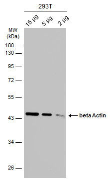
- Experimental details
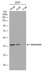
- Various whole cell extracts (15 | 5 | 2 ?g) were separated by 10% SDS-PAGE, and the membrane was blotted with beta Actin antibody (GTX110564) diluted at 1:10000. The HRP-conjugated anti-rabbit IgG antibody (GTX213110-01) was used to detect the primary antibody.
- Submitted by
- GeneTex (provider)
- Main image
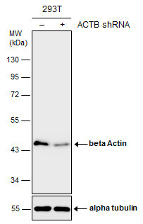
- Experimental details
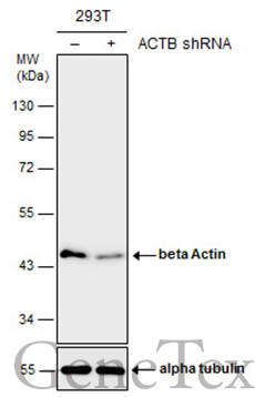
- Non-transfected (¡V) and transfected (+) 293T whole cell extracts (10 ?g) were separated by 10% SDS-PAGE, and the membrane was blotted with beta Actin antibody (GTX110564) diluted at 1:15000. The HRP-conjugated anti-rabbit IgG antibody (GTX213110-01) was used to detect the primary antibody.
- Submitted by
- GeneTex (provider)
- Main image
- Experimental details
- Various whole cell extracts were separated by 10% SDS-PAGE, and the membrane was blotted with beta Actin antibody (GTX109639) diluted at 1:10000.

- Submitted by
- GeneTex (provider)
- Main image

- Experimental details
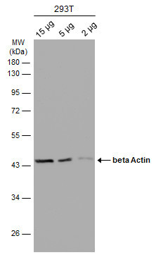
- Various whole cell extracts (15 | 5 | 2 ?g) were separated by 10% SDS-PAGE, and the membrane was blotted with beta Actin antibody (GTX110564) diluted at 1:10000. The HRP-conjugated anti-rabbit IgG antibody (GTX213110-01) was used to detect the primary antibody.
- Submitted by
- GeneTex (provider)
- Main image
- Experimental details
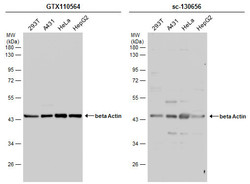
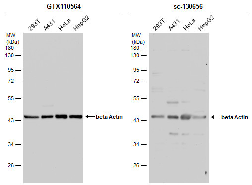
- Various whole cell extracts (30 ?g) were separated by 10% SDS-PAGE, and the membranes were blotted with beta Actin antibody (GTX110564) diluted at 1:10000 and competitor's antibody (sc-130656) diluted at 1:100. The HRP-conjugated anti-rabbit IgG antibody (GTX213110-01) was used to detect the primary antibody.
Supportive validation
- Submitted by
- GeneTex (provider)
- Main image
- Experimental details
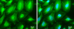
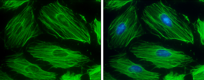
- beta Actin antibody detects beta Actin protein at cytoplasm and cytoskeleton by immunofluorescent analysis.Sample: HeLa cells were fixed in ice-cold MeOH for 5 min.Green: beta Actin protein stained by beta Actin antibody (GTX110564) diluted at 1:500.Blue: Hoechst 33342 staining.Scale bar = 10 £gm.
- Submitted by
- GeneTex (provider)
- Main image

- Experimental details
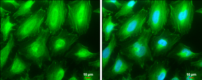
- beta Actin antibody detects beta Actin protein at cytoskeleton by immunofluorescent analysis.Sample: HeLa cells were fixed in 0.5% Triton X-100 for 1 min, then ice-cold methanol for 5 min.Green: beta Actin protein stained by beta Actin antibody (GTX110564) diluted at 1:500.Blue: Hoechst 33342 staining.
Supportive validation
- Submitted by
- GeneTex (provider)
- Main image
- Experimental details
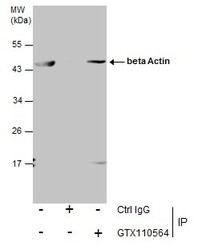
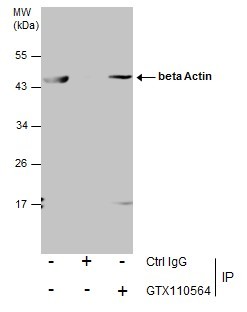
- Immunoprecipitation of beta Actin protein from 293T whole cell extracts using 5 £gg of beta Actin antibody (GTX110564).Western blot analysis was performed using beta Actin antibody (GTX110564).EasyBlot anti-Rabbit IgG (GTX221666-01) was used as a secondary reagent.
Supportive validation
- Submitted by
- GeneTex (provider)
- Main image
- Experimental details
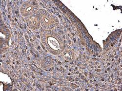
- beta Actin antibody detects beta Actin protein at cytoplasm in mouse cervix by immunohistochemical analysis. Sample: Paraffin-embedded mouse cervix. beta Actin antibody (GTX110564) diluted at 1:500.

- Submitted by
- GeneTex (provider)
- Main image
- Experimental details
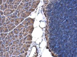
- beta Actin antibody detects beta Actin protein at cell membrane and cytoplasm in mouse pancreas by immunohistochemical analysis. Sample: Paraffin-embedded mouse pancreas. beta Actin antibody (GTX110564) diluted at 1:500.
- Submitted by
- GeneTex (provider)
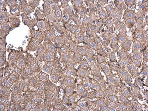
- Main image

- Experimental details
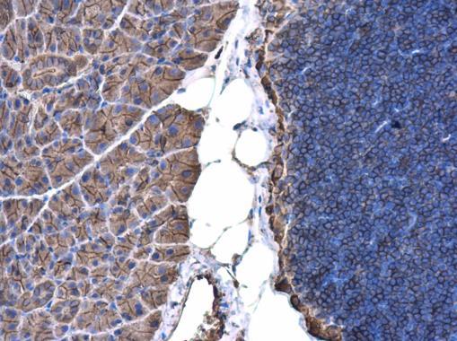
- beta Actin antibody detects beta Actin protein at cell membrane and cytoplasm in rat liver by immunohistochemical analysis. Sample: Paraffin-embedded rat liver. beta Actin antibody (GTX110564) diluted at 1:500.