Explore
 Validate
 Learn
Learn Western blot
Antibody data
- Antibody Data
- Antigen structure
- References [0]
- Comments [0]
- Validations
- Western blot [4]
- Immunocytochemistry [2]
- Immunohistochemistry [11]
Submit
Validation data
Reference
Comment
Report error
- Product number
- GTX124212 - Provider product page

- Provider
- GeneTex
- Proper citation
- GeneTex Cat#GTX124212, RRID:AB_11170726
- Product name
- beta Actin antibody
- Antibody type
- Polyclonal
- Reactivity
- Human, Mouse, Rat
- Host
- Rabbit
No comments: Submit comment
Supportive validation
- Submitted by
- GeneTex (provider)
- Main image

- Experimental details
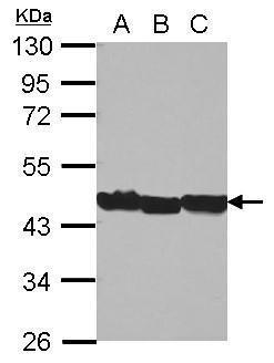
- Sample (30 ug of whole cell lysate) A: Jurkat B: Raji C: THP-1 10% SDS PAGE GTX124212 diluted at 1:10000
- Submitted by
- GeneTex (provider)
- Main image

- Experimental details
- Sample (30 ug of whole cell lysate) A: NIH-3T3 10% SDS PAGE GTX124212 diluted at 1:10000
- Submitted by
- GeneTex (provider)
- Main image

- Experimental details
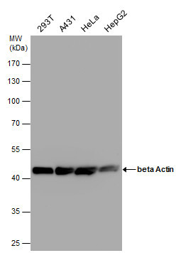
- beta Actin antibody detects beta Actin protein by western blot analysis. Various whole cell extracts (30 £gg) were separated by 10% SDS-PAGE, and the membrane was blotted with beta Actin antibody (GTX124212) diluted by 1:10000.
- Submitted by
- GeneTex (provider)
- Main image

- Experimental details
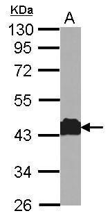
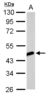
- Beta Actin antibody detects beta Actin protein by Western blot analysis.A. 1 £gg 293T whole cell lysate/extract10 % SDS-PAGEbeta Actin antibody (GTX124212) dilution: 1:10000
Supportive validation
- Submitted by
- GeneTex (provider)
- Main image
- Experimental details
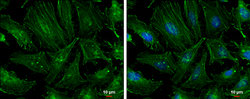
- beta Actin antibody detects beta Actin protein at cytoskeleton by immunofluorescent analysis.Sample: HeLa cells were fixed in ice-cold MeOH for 5 min.Green: beta Actin protein stained by beta Actin antibody (GTX124212) diluted at 1:500.Blue: Hoechst 33342 staining.Scale bar = 10 £gm.
- Submitted by
- GeneTex (provider)
- Main image

- Experimental details
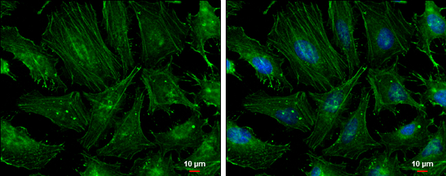
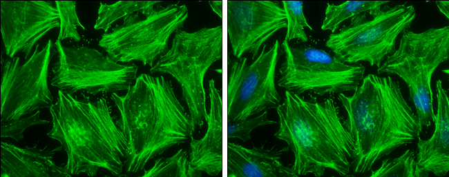
- beta Actin antibody detects beta Actin protein at cytoskeleton by immunofluorescent analysis.Sample: HeLa cells were fixed in ice-cold MeOH for 5 min.Green: beta Actin protein stained by beta Actin antibody (GTX124212) diluted at 1:500.Blue: Hoechst 33342 staining.
Supportive validation
- Submitted by
- GeneTex (provider)
- Main image

- Experimental details
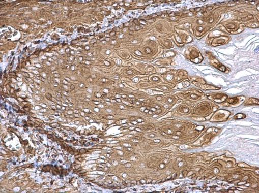
- beta Actin antibody detects beta Actin protein at cytosol on mouse middle brain by immunohistochemical analysis. Sample: Paraffin-embedded mouse esophagus. beta Actin antibody (GTX124212) dilution: 1:500.
- Submitted by
- GeneTex (provider)
- Main image

- Experimental details
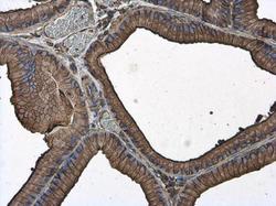
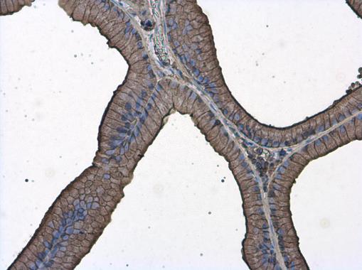
- Beta Actin antibody detects beta Actin protein at cytoplasm in rat prostate by immunohistochemical analysis. Sample: Paraffin-embedded rat prostate. Beta Actin antibody (GTX124212) diluted at 1:500.

- Submitted by
- GeneTex (provider)
- Main image

- Experimental details
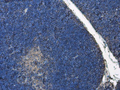
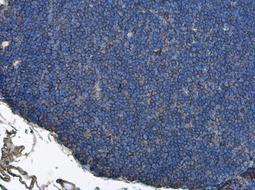
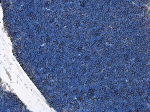
- Beta Actin antibody detects beta Actin protein at cytoplasm in rat thymus gland by immunohistochemical analysis. Sample: Paraffin-embedded rat thymus gland. Beta Actin antibody (GTX124212) diluted at 1:500.
- Submitted by
- GeneTex (provider)
- Main image
- Experimental details
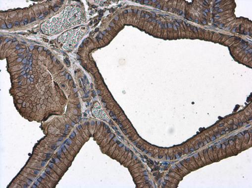
- Beta Actin antibody detects beta Actin protein at cytoplasm in rat prostate by immunohistochemical analysis. Sample: Paraffin-embedded rat prostate. Beta Actin antibody (GTX124212) diluted at 1:500.
- Submitted by
- GeneTex (provider)
- Main image

- Experimental details
- Beta Actin antibody detects beta Actin protein at cytoplasm in mouse thymus gland by immunohistochemical analysis. Sample: Paraffin-embedded mouse thymus gland. Beta Actin antibody (GTX124212) diluted at 1:500.
- Submitted by
- GeneTex (provider)
- Main image

- Experimental details
- Beta Actin antibody detects beta Actin protein at cytoplasm in rat prostate by immunohistochemical analysis. Sample: Paraffin-embedded rat prostate. Beta Actin antibody (GTX124212) diluted at 1:500.
- Submitted by
- GeneTex (provider)
- Main image
- Experimental details
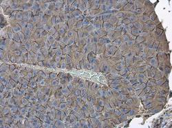
- Beta Actin antibody detects beta Actin protein at cytoplasm in mouse pancreas by immunohistochemical analysis. Sample: Paraffin-embedded mouse pancreas. Beta Actin antibody (GTX124212) diluted at 1:500.
- Submitted by
- GeneTex (provider)
- Main image

- Experimental details
- Beta Actin antibody detects beta Actin protein at cytoplasm in rat thymus gland by immunohistochemical analysis. Sample: Paraffin-embedded rat thymus gland. Beta Actin antibody (GTX124212) diluted at 1:500.
- Submitted by
- GeneTex (provider)
- Main image

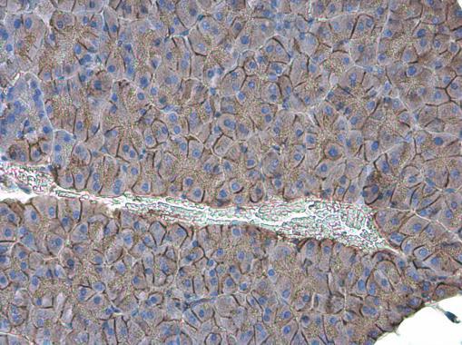
- Experimental details
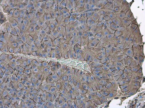
- Beta Actin antibody detects beta Actin protein at cytoplasm in mouse pancreas by immunohistochemical analysis. Sample: Paraffin-embedded mouse pancreas. Beta Actin antibody (GTX124212) diluted at 1:500.
- Submitted by
- GeneTex (provider)
- Main image

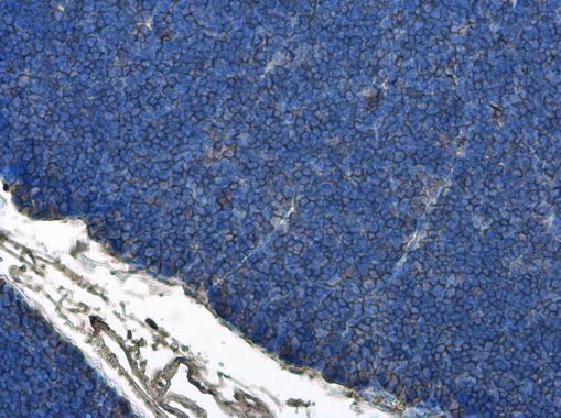
- Experimental details
- Beta Actin antibody detects beta Actin protein at cytoplasm in mouse thymus gland by immunohistochemical analysis. Sample: Paraffin-embedded mouse thymus gland. Beta Actin antibody (GTX124212) diluted at 1:500.
- Submitted by
- GeneTex (provider)
- Main image

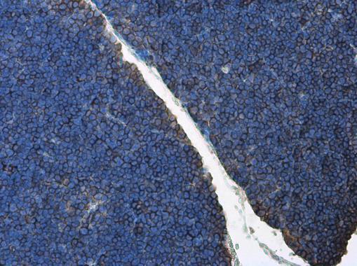
- Experimental details
- Beta Actin antibody detects beta Actin protein at cytoplasm in mouse thymus gland by immunohistochemical analysis. Sample: Paraffin-embedded mouse thymus gland. Beta Actin antibody (GTX124212) diluted at 1:500.