 Explore
 Validate
 Learn
Learn Western blot
 Immunocytochemistry
Antibody data
- Antibody Data
- Antigen structure
- References [3]
- Comments [0]
- Validations
- Western blot [4]
- Immunocytochemistry [1]
- Immunohistochemistry [2]
Submit
Validation data
Reference
Comment
Report error
- Product number
- GTX124214 - Provider product page

- Provider
- GeneTex
- Proper citation
- GeneTex Cat#GTX124214, RRID:AB_11177721
- Product name
- beta Actin antibody
- Antibody type
- Polyclonal
- Reactivity
- Human, Mouse
- Host
- Rabbit
Submitted references Linear mitochondrial DNA is rapidly degraded by components of the replication machinery.
Tunable and reversible drug control of protein production via a self-excising degron.
In vivo quantitative proteomics of somatosensory cortical synapses shows which protein levels are modulated by sensory deprivation.
Peeva V, Blei D, Trombly G, Corsi S, Szukszto MJ, Rebelo-Guiomar P, Gammage PA, Kudin AP, Becker C, Altmüller J, Minczuk M, Zsurka G, Kunz WS
Nature communications 2018 Apr 30;9(1):1727
Nature communications 2018 Apr 30;9(1):1727
Tunable and reversible drug control of protein production via a self-excising degron.
Chung HK, Jacobs CL, Huo Y, Yang J, Krumm SA, Plemper RK, Tsien RY, Lin MZ
Nature chemical biology 2015 Sep;11(9):713-20
Nature chemical biology 2015 Sep;11(9):713-20
In vivo quantitative proteomics of somatosensory cortical synapses shows which protein levels are modulated by sensory deprivation.
Butko MT, Savas JN, Friedman B, Delahunty C, Ebner F, Yates JR 3rd, Tsien RY
Proceedings of the National Academy of Sciences of the United States of America 2013 Feb 19;110(8):E726-35
Proceedings of the National Academy of Sciences of the United States of America 2013 Feb 19;110(8):E726-35
No comments: Submit comment
Enhanced validation
Supportive validation
- Submitted by
- GeneTex (provider)
- Enhanced method
- Genetic validation
- Main image

- Experimental details
- Non-transfected (¡V) and transfected (+) 293T whole cell extracts (10 ?g) were separated by 10% SDS-PAGE, and the membrane was blotted with beta Actin antibody (GTX124214) diluted at 1:15000.

Supportive validation
- Submitted by
- GeneTex (provider)
- Main image

- Experimental details
- Sample (30 ug of whole cell lysate) A: NIH-3T3 10% SDS PAGE GTX124214 diluted at 1:10000

- Submitted by
- GeneTex (provider)
- Main image

- Experimental details
- Sample (30 ug of whole cell lysate) A: Jurakt B: Raji C: K562 10% SDS PAGE GTX124214 diluted at 1:10000

- Submitted by
- GeneTex (provider)
- Main image

- Experimental details
- Non-transfected (¡V) and transfected (+) 293T whole cell extracts (10 ?g) were separated by 10% SDS-PAGE, and the membrane was blotted with beta Actin antibody (GTX124214) diluted at 1:15000.

Supportive validation
- Submitted by
- GeneTex (provider)
- Main image

- Experimental details
- Immunofluorescence analysis of methanol-fixed HeLa, using beta actin(GTX124214) antibody at 1:500 dilution.

Supportive validation
- Submitted by
- GeneTex (provider)
- Main image
- Experimental details
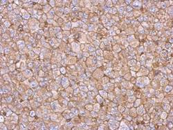
- Immunohistochemical analysis of paraffin-embedded H1299 xenograft, using beta actin(GTX124214) antibody at 1:500 dilution.

- Submitted by
- GeneTex (provider)
- Main image
- Experimental details
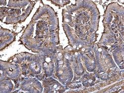
- beta actin antibody detects beta actin protein at cytosol on mouse intestine by immunohistochemical analysis. Sample: Paraffin-embedded mouse intestine. beta actin antibody (GTX124214) dilution: 1:500.
