Explore
 Validate
 Learn
Learn Western blot
Antibody data
- Antibody Data
- Antigen structure
- References [0]
- Comments [0]
- Validations
- Western blot [2]
- Immunocytochemistry [1]
Submit
Validation data
Reference
Comment
Report error
- Product number
- MA1-140-A555 - Provider product page

- Provider
- Invitrogen Antibodies
- Product name
- beta Actin Monoclonal Antibody (15G5A11/E2), Alexa Fluor™ 555
- Antibody type
- Monoclonal
- Antigen
- Purifed from natural sources
- Reactivity
- Human, Mouse, Rat
- Host
- Mouse
- Isotype
- IgG
- Antibody clone number
- 15G5A11/E2
- Vial size
- 50 µL
- Concentration
- 1 mg/mL
- Storage
- 4° C, do not freeze
No comments: Submit comment
Supportive validation
- Submitted by
- Invitrogen Antibodies (provider)
- Main image
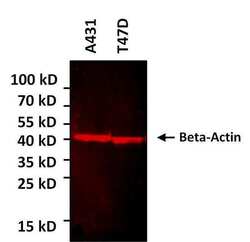
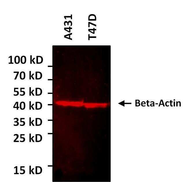
- Experimental details
- Western blot analysis of beta-Actin was performed by loading various cell lysates onto a 4-20% Tris-HCl polyacrylamide gel. Proteins were transferred to a low fluorescence PVDF membrane and blocked with Sea Block blocking buffer for at least 1 hour. The membrane was probed with a AlexaFluor555-conjugated beta-Actin monoclonal antibody (Product # MA1-140-A555) at a dilution of 1:500 for over night at 4C on a rocking platform and washed in TBS-0.1% Tween-20. Detection was performed using a fluorescence imaging system.
- Submitted by
- Invitrogen Antibodies (provider)
- Main image

- Experimental details
- Western Blot was performed using Anti-beta Actin Monoclonal Antibody (15G5A11/E2), Alexa Fluor 555 (Product # MA1-140-A555) and a ~40 kDa band corresponding to Actin, cytoplasmic 1 was observed along with an uncharacterised band (*) at ~30 kDa across cell lines and tissues tested. Whole cell extracts (30 µg lysate) of HeLa (Lane 1), A549 (Lane 2), A-431 (Lane 3), U-2 OS (Lane 4), Jurkat (Lane 5), PC-12 (Lane 6), NIH/3T3 (Lane 7), Mouse Spleen (Lane 8), Mouse Liver (Lane 9), Rat Spleen (Lane 10), Rat Liver (Lane 11) were electrophoresed using NuPAGE™ 4-12% Bis-Tris Protein Gel (Product # NP0321BOX). Resolved proteins were then transferred onto a Nitrocellulose membrane (Product # LC2001) by iBlot® 2 Dry Blotting System (Product # IB21001). The Blot was probed with the primary antibody (1:1000 dilution) and detected by fluorescence using the iBright FL 1000 (Product # A32752). Fluorescent detection was performed. doi:10.1038/bcj.2014.18

Supportive validation
- Submitted by
- Invitrogen Antibodies (provider)
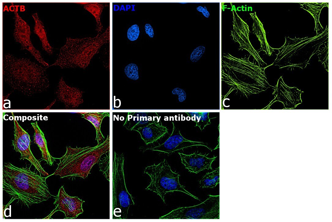
- Main image

- Experimental details
- Immunofluorescence analysis of Actin, cytoplasmic 1 was performed using 70% confluent log phase HeLa cells. The cells were fixed with 4% paraformaldehyde for 5 minutes, permeabilized with 0.1% Triton™ X-100 for 10 minutes, and blocked with 2% BSA for 5 minutes at room temperature. The cells were labeled with beta Actin Monoclonal Antibody (15G5A11/E2), Alexa Fluor 555 (Product # MA1-140-A555) at 1:100 dilution in 0.1% BSA, incubated at 4 degree celsius overnight (Panel a: Red). Nuclei (Panel b:Blue) were stained with ProLong™ Diamond Antifade Mountant with DAPI (Product # P36962). F-actin (Panel c: Green) was stained with Alexa Fluor™ 488 Phalloidin (Product # A12379, 1:300). Panel d represents the merged image showing Cytoskeleton localization. Panel e represents control cells with no primary antibody to assess background. The images were captured at 60x magnification.