 Explore
 Validate
 Learn
Learn Western blot
Antibody data
- Antibody Data
- Antigen structure
- References [4]
- Comments [0]
- Validations
- Western blot [1]
- Other assay [5]
Submit
Validation data
Reference
Comment
Report error
- Product number
- PA1-183-HRP - Provider product page

- Provider
- Invitrogen Antibodies
- Product name
- beta Actin Polyclonal Antibody, HRP
- Antibody type
- Polyclonal
- Antigen
- Purifed from natural sources
- Description
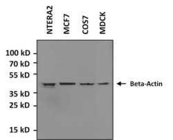
- By Western blot, PA1-183-HRP detects a prominent ~42kD protein in human, mouse, non-human primate, and rat whole cell lysates.
- Reactivity
- Human, Mouse, Rat
- Host
- Rabbit
- Conjugate
- Horseradish Peroxidase
- Isotype
- IgG
- Vial size
- 50 µL
- Concentration
- 1 mg/mL
- Storage
- 4° C, do not freeze
Submitted references LncRNA ASAP1-IT1 enhances cancer cell stemness via regulating miR-509-3p/YAP1 axis in NSCLC.
PIK3CA Is Regulated by CUX1, Promotes Cell Growth and Metastasis in Bladder Cancer via Activating Epithelial-Mesenchymal Transition.
Replication and ribosomal stress induced by targeting pyrimidine synthesis and cellular checkpoints suppress p53-deficient tumors.
A point mutation decouples the lipid transfer activities of microsomal triglyceride transfer protein.
Liu Y, Yang Y, Zhang L, Lin J, Li B, Yang M, Li H, Chen K, Zhao W
Cancer cell international 2021 Oct 29;21(1):572
Cancer cell international 2021 Oct 29;21(1):572
PIK3CA Is Regulated by CUX1, Promotes Cell Growth and Metastasis in Bladder Cancer via Activating Epithelial-Mesenchymal Transition.
Wang Z, Shang J, Li Z, Li H, Zhang C, He K, Li S, Ju W
Frontiers in oncology 2020;10:536072
Frontiers in oncology 2020;10:536072
Replication and ribosomal stress induced by targeting pyrimidine synthesis and cellular checkpoints suppress p53-deficient tumors.
Hubackova S, Davidova E, Boukalova S, Kovarova J, Bajzikova M, Coelho A, Terp MG, Ditzel HJ, Rohlena J, Neuzil J
Cell death & disease 2020 Feb 7;11(2):110
Cell death & disease 2020 Feb 7;11(2):110
A point mutation decouples the lipid transfer activities of microsomal triglyceride transfer protein.
Wilson MH, Rajan S, Danoff A, White RJ, Hensley MR, Quinlivan VH, Recacha R, Thierer JH, Tan FJ, Busch-Nentwich EM, Ruddock L, Hussain MM, Farber SA
PLoS genetics 2020 Aug;16(8):e1008941
PLoS genetics 2020 Aug;16(8):e1008941
No comments: Submit comment
Supportive validation
- Submitted by
- Invitrogen Antibodies (provider)
- Main image
- Experimental details
- Western blot analysis of beta-Actin was performed by loading various whole cell lysates onto a 4-20% Tris-HCl polyacrylamide gel. Proteins were transferred to a PVDF membrane and blocked with 5% BSA/TBST for at least 1 hour. Membranes were probed with beta-Actin polyclonal antibody (Product # PA1-183-HRP) at a dilution of 1:500 for 1 hour at room temperature on a rocking platform and washed in TBS-0.1% Tween-20. Chemiluminescent detection was performed using Super Signal West Pico (Product # 34080).
- Conjugate
- Horseradish Peroxidase

Supportive validation
- Submitted by
- Invitrogen Antibodies (provider)
- Main image

- Experimental details
- NULL
- Conjugate
- Horseradish Peroxidase

- Submitted by
- Invitrogen Antibodies (provider)
- Main image

- Experimental details
- NULL
- Conjugate
- Horseradish Peroxidase

- Submitted by
- Invitrogen Antibodies (provider)
- Main image

- Experimental details
- Fig. 6 YAP1 is regulated by miR-509-3p and ASAP1-IT1. A The potential target of miR-509-3p is predicted by TargetScan. B qRT-PCR analysis of YAP1 expression in NSCLC tissues and adjacent tissues, * p < 0.01 compared with adjacent tissues. C qRT-PCR analysis of YAP1 in A549 spheres, * p < 0.01 compared with parental group. D Luciferase activity of pmirGLO-YAP1-3'UTR-WT (wild type) or pmirGLO-YAP1-3'UTR-Mut (mutant) was modulated by miR-509-3p and ASAP1-IT1 in A549 cells, * p < 0.01 compared with Mock + pcDNA, phi p < 0.01 compared with miR-509-3p + pcDNA, F p < 0.01 compared with miR-509-3p + ASAP1-IT1. E YAP1 mRNA levels in A549 cells analyzed byqRT-PCR, * p < 0.01 compared with Mock + pcDNA, phi p < 0.01 compared with miR-509-3p + pcDNA, F p < 0.01 compared with miR-509-3p + ASAP1-IT1. F Western blot analysis on YAP1 protein in A549 cells
- Conjugate
- Horseradish Peroxidase

- Submitted by
- Invitrogen Antibodies (provider)
- Main image

- Experimental details
- Fig. 2 Inhibition of DHODH stabilizes the p53 tumor suppressor protein. a Parental 4T1 cells and D0-D25 sublines were subjected to microchip analysis and a heat map for the 119 transcripts involved in ribosome biogenesis was constructed. b - e , g MCF7 and HCT116 cells were exposed to leflunomide (LFM, 50 uM) for 72 h in the presence or absence of uridine (U; 50 ug/l). Expression of 45S rRNA ( b ) and 18S rRNA ( c ) was assessed by qRT-PCR. d Immunoblotting detection of RPS6 was performed. beta-Actin was used as a loading control. e Immunofluorescent detection of B23 in MCF7 cells was performed. f Immunoblotting detection of p53 pS15, p53, and p21 in MCF7 cells treated 72 h with LFM (50 uM) after downregulation of RPL5 and RPL11 alone or in combination using specific siRNA. Non-targeting siRNA (siNC) was used as a control. beta-Actin was used as a loading control. g Immunoblot detection of MDM2 in MCF7 and HCT116 cells. beta-Actin was used as a loading control. h Co-immunoprecipitation of p53 followed by immunoblot analysis of p53, MDM2, ubiquitinated p53, and ubiquitinated MDM2 in MCF7 control and LFM (50 uM, 72 h)-treated cells. GAPDH was used as an input control. Immunoprecipitated samples and input represent one membrane with different intensity of signal. i Levels of ubiquitinated p53 and MDM2 related to their immunoprecipitated total forms were evaluated from three independent experiments. j Co-immunoprecipitation of MDM2 followed by immunoblot analysi
- Conjugate
- Horseradish Peroxidase

- Submitted by
- Invitrogen Antibodies (provider)
- Main image

- Experimental details
- Fig. 3 Role of p53 in cell cycle arrest after leflunomide treatment. a MCF7 and b HCT116 cells were exposed to LFM (50 uM) for 72 h after downregulation of RPL5 and RPL11 alone or in combination using specific siRNA. Cell death was assessed by flow cytometry using annexin V/Hoechst staining. Non-targeting siRNA (siNC) was used as a control. c MCF7 cells were transfected with specific p53 siRNA or with non-targeting siRNA (siNC), respectively. Cell death was evaluated by annexin V/Hoechst positivity using FACS. Immunoblot detection of p53 and p21 shows the effect of transfection. beta-Actin was used as a loading control. d HCT116 wt p53 and HCT116 p53 KO cells were exposed to LFM (50 uM) for 72 h and cell death was determined by flow cytometry using annexin V/Hoechst staining. Immunoblot detection of p53 and p21 show the effect of p53 KO . beta-Actin was used as a loading control. In a - d , data are shown as mean +- SEM, n = 3-5. * P < 0.05, two-way ANOVA.
- Conjugate
- Horseradish Peroxidase
