Explore
 Validate
 Learn
Learn Western blot
 ELISA
ELISAAntibody data
- Antibody Data
- Antigen structure
- References [9]
- Comments [0]
- Validations
- Western blot [4]
- Immunocytochemistry [3]
- Flow cytometry [1]
- Other assay [4]
Submit
Validation data
Reference
Comment
Report error
- Product number
- MA5-15452 - Provider product page

- Provider
- Invitrogen Antibodies
- Product name
- beta Actin Monoclonal Antibody (8H10D10)
- Antibody type
- Monoclonal
- Antigen
- Synthetic peptide
- Description
- MA5-15452 targets beta-Actin in indirect ELISA, FACS, IF, and WB applications and shows reactivity with Hamster, Human, mouse, Non-human primate, and Rat samples. The MA5-15452 immunogen is synthetic peptide corresponding to amino-terminal residues of human beta-Actin, conjugated to KLH. MA5-15452 detects beta-Actin which has a predicted molecular weight of approximately 42kDa.
- Reactivity
- Human, Mouse, Rat, Hamster
- Host
- Mouse
- Isotype
- IgG
- Antibody clone number
- 8H10D10
- Vial size
- 100 µL
- Concentration
- Conc. Not Determined
- Storage
- Store at 4°C short term. For long term storage, store at -20°C, avoiding freeze/thaw cycles.
Submitted references Parecoxib inhibits esophageal squamous cell carcinoma progression via the PDK1-AKT pathway.
Low-Dose Albendazole Inhibits Epithelial-Mesenchymal Transition of Melanoma Cells by Enhancing Phosphorylated GSK-3β/Tyr216 Accumulation.
The hepcidin regulator erythroferrone is a new member of the erythropoiesis-iron-bone circuitry.
Immunomodulatory effects of Radix isatidis polysaccharides in vitro and in vivo.
Cathepsin S Regulates Antigen Processing and T Cell Activity in Non-Hodgkin Lymphoma.
Prohibitin is a prognostic marker and therapeutic target to block chemotherapy resistance in Wilms' tumor.
Chromatin Remodeling in Response to BRCA2-Crisis.
Influence of Matrices on 3D-Cultured Prostate Cancer Cells' Drug Response and Expression of Drug-Action Associated Proteins.
An Antibody-Drug Conjugate Directed against Lymphocyte Antigen 6 Complex, Locus E (LY6E) Provides Robust Tumor Killing in a Wide Range of Solid Tumor Malignancies.
Huang HM, Huang XY, Wu SP, Chen CK, He XH, Zhang YF
Cellular & molecular biology letters 2022 Mar 19;27(1):28
Cellular & molecular biology letters 2022 Mar 19;27(1):28
Low-Dose Albendazole Inhibits Epithelial-Mesenchymal Transition of Melanoma Cells by Enhancing Phosphorylated GSK-3β/Tyr216 Accumulation.
He Z, Lei S, Liang F, Tan L, Zhang W, Xie L, Zheng H, Lu Y
Journal of oncology 2021;2021:4475192
Journal of oncology 2021;2021:4475192
The hepcidin regulator erythroferrone is a new member of the erythropoiesis-iron-bone circuitry.
Castro-Mollo M, Gera S, Ruiz-Martinez M, Feola M, Gumerova A, Planoutene M, Clementelli C, Sangkhae V, Casu C, Kim SM, Ostland V, Han H, Nemeth E, Fleming R, Rivella S, Lizneva D, Yuen T, Zaidi M, Ginzburg Y
eLife 2021 May 18;10
eLife 2021 May 18;10
Immunomodulatory effects of Radix isatidis polysaccharides in vitro and in vivo.
Tao W, Fu T, He ZJ, Zhou HP, Hong Y
Experimental and therapeutic medicine 2021 Dec;22(6):1405
Experimental and therapeutic medicine 2021 Dec;22(6):1405
Cathepsin S Regulates Antigen Processing and T Cell Activity in Non-Hodgkin Lymphoma.
Dheilly E, Battistello E, Katanayeva N, Sungalee S, Michaux J, Duns G, Wehrle S, Sordet-Dessimoz J, Mina M, Racle J, Farinha P, Coukos G, Gfeller D, Mottok A, Kridel R, Correia BE, Steidl C, Bassani-Sternberg M, Ciriello G, Zoete V, Oricchio E
Cancer cell 2020 May 11;37(5):674-689.e12
Cancer cell 2020 May 11;37(5):674-689.e12
Prohibitin is a prognostic marker and therapeutic target to block chemotherapy resistance in Wilms' tumor.
Ortiz MV, Ahmed S, Burns M, Henssen AG, Hollmann TJ, MacArthur I, Gunasekera S, Gaewsky L, Bradwin G, Ryan J, Letai A, He Y, Naranjo A, Chi YY, LaQuaglia M, Heaton T, Cifani P, Dome JS, Gadd S, Perlman E, Mullen E, Steen H, Kentsis A
JCI insight 2019 Aug 8;4(15)
JCI insight 2019 Aug 8;4(15)
Chromatin Remodeling in Response to BRCA2-Crisis.
Gruber JJ, Chen J, Geller B, Jäger N, Lipchik AM, Wang G, Kurian AW, Ford JM, Snyder MP
Cell reports 2019 Aug 20;28(8):2182-2193.e6
Cell reports 2019 Aug 20;28(8):2182-2193.e6
Influence of Matrices on 3D-Cultured Prostate Cancer Cells' Drug Response and Expression of Drug-Action Associated Proteins.
Edmondson R, Adcock AF, Yang L
PloS one 2016;11(6):e0158116
PloS one 2016;11(6):e0158116
An Antibody-Drug Conjugate Directed against Lymphocyte Antigen 6 Complex, Locus E (LY6E) Provides Robust Tumor Killing in a Wide Range of Solid Tumor Malignancies.
Asundi J, Crocker L, Tremayne J, Chang P, Sakanaka C, Tanguay J, Spencer S, Chalasani S, Luis E, Gascoigne K, Desai R, Raja R, Friedman BA, Haverty PM, Polakis P, Firestein R
Clinical cancer research : an official journal of the American Association for Cancer Research 2015 Jul 15;21(14):3252-62
Clinical cancer research : an official journal of the American Association for Cancer Research 2015 Jul 15;21(14):3252-62
No comments: Submit comment
Supportive validation
- Submitted by
- Invitrogen Antibodies (provider)
- Main image

- Experimental details
- Western blot analysis of beta-Actin using beta-Actin monoclonal antibody (Product # MA5-15452) in NIH/3T3 (1), Jurkat (2), HeLa (3), CHO (4), PC12 (5), HEK293 (6), COS-7 (7), A549 (8) and MCF-7 (9) cell lysate.

- Submitted by
- Invitrogen Antibodies (provider)
- Main image

- Experimental details
- Western blot analysis of beta-Actin using beta-Actin monoclonal antibody (Product # MA5-15452) in NIH/3T3 (1), Jurkat (2), HeLa (3), CHO (4), PC12 (5), HEK293 (6), COS-7 (7), A549 (8) and MCF-7 (9) cell lysate.

- Submitted by
- Invitrogen Antibodies (provider)
- Main image

- Experimental details
- Western blot analysis of beta-Actin using beta-Actin monoclonal antibody (Product # MA5-15452) in NIH/3T3 (1), Jurkat (2), HeLa (3), CHO (4), PC12 (5), HEK293 (6), COS-7 (7), A549 (8) and MCF-7 (9) cell lysate.

- Submitted by
- Invitrogen Antibodies (provider)
- Main image

- Experimental details
- Western Blot was performed using Anti-beta Actin Monoclonal Antibody (8H10D10) (Product # MA5-15452) and a ~40 kDa band corresponding to Actin, cytoplasmic 1 was observed across cell lines and tissues tested. Whole cell extracts (30 µg lysate) of HeLa (Lane 1), A549 (Lane 2), A-431 (Lane 3), U-2 OS (Lane 4), Jurkat (Lane 5), PC-12 (Lane 6), NIH/3T3 (Lane 7), Mouse Spleen (Lane 8), Mouse Liver (Lane 9), Rat Spleen (Lane 10), Rat Liver (Lane 11) were electrophoresed using NuPAGE™ 4-12% Bis-Tris Protein Gel (Product # NP0321BOX). Resolved proteins were then transferred onto a Nitrocellulose membrane (Product # LC2001) by iBlot® 2 Dry Blotting System (Product # IB21001). The Blot was probed with the primary antibody (1:2000 dilution) and detected by chemiluminescence with Goat anti-Mouse IgG (H+L) Superclonal™ Recombinant Secondary Antibody, HRP (Product # A28177, 1:4000 dilution) using the iBright FL 1000 (Product # A32752). Chemiluminescent detection was performed using Novex® ECL Chemiluminescent Substrate Reagent Kit (Product # WP20005). doi:10.1038/bcj.2014.18

Supportive validation
- Submitted by
- Invitrogen Antibodies (provider)
- Main image
- Experimental details
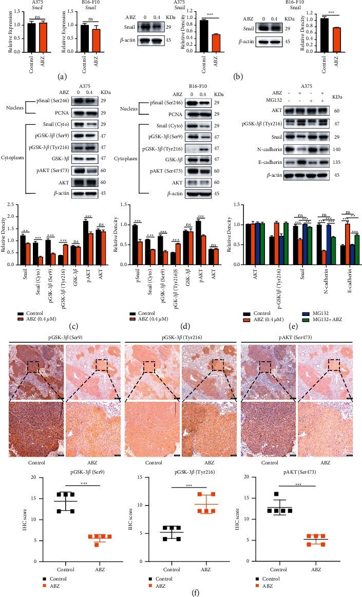
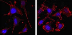
- Immunofluorescence analysis of SKBR-3 (left) and A549 (right) cells using beta-Actin monoclonal antibody (Product # MA5-15452) (Red). Blue: DRAQ5 fluorescent DNA dye. Red: the secondary Ab is Cy3-Goat anti mouse IgG

- Submitted by
- Invitrogen Antibodies (provider)
- Main image
- Experimental details
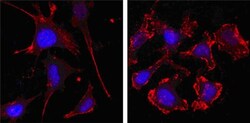
- Immunofluorescence analysis of Actin, cytoplasmic 1 was performed using 70% confluent log phase HeLa cells. The cells were fixed with 4% paraformaldehyde for 5 minutes, permeabilized with 0.1% Triton™ X-100 for 10 minutes, and blocked with 2% BSA for 45 minutes at room temperature. The cells were labeled with beta Actin Monoclonal Antibody (8H10D10) (Product # MA5-15452) at 1:100 dilution in 0.1% BSA, incubated at 4 degree celsius overnight and then labeled with Donkey anti-Mouse IgG (H+L) Highly Cross-Adsorbed Secondary Antibody, Alexa Fluor Plus 647 (Product # A32787), (1:2000 dilution), for 45 minutes at room temperature (Panel a: Green). Nuclei (Panel b:Blue) were stained with ProLong™ Diamond Antifade Mountant with DAPI (Product # P36962). F-actin (Panel c: Red) was stained with Rhodamine Phalloidin (Product # R415, 1:300). Panel d represents the merged image showing Cytoskeleton localization. Panel e represents control cells with no primary antibody to assess background. The images were captured at 60x magnification.

Supportive validation
- Submitted by
- Invitrogen Antibodies (provider)
- Main image

- Experimental details
- Flow cytometric analysis of MCF-7 cells using beta-Actin monoclonal antibody (Product # MA5-15452) (right) and negative control (left).

Supportive validation
- Submitted by
- Invitrogen Antibodies (provider)
- Main image

- Experimental details
- NULL

- Submitted by
- Invitrogen Antibodies (provider)
- Main image

- Experimental details
- NULL

- Submitted by
- Invitrogen Antibodies (provider)
- Main image

- Experimental details
- NULL

- Submitted by
- Invitrogen Antibodies (provider)
- Main image
- Experimental details
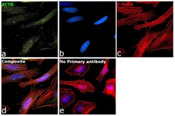
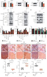
- Figure 4 ABZ treatment downregulates the snail expression in melanoma cells by increasing the accumulation of phosphorylated GSK-3 beta /Tyr216. (a) The relative transcription levels of Snail in the ABZ-treated (0.4 mu M) and control groups of A375 (left) and B16-F10 (right) melanoma cells were measured by RT-qPCR, with beta -actin as the internal control. (b) The expression of transcription factor Snail in A375 (left) and B16-F10 (right) cells was detected by western blot analysis, with beta -actin as the internal reference protein. (c-d) The expression levels of cytoplasmic proteins AKT, pAKT, GSK-3 beta , pGSK-3 beta (Ser9/Tyr216) and Snail, and nuclear protein pSnail in A375 and B16-F10 cells were also determined by western blotting, with beta -actin and PCNA as the internal controls for the cytoplasmic and nuclear proteins, respectively. The histograms show the relative density of AKT/pAKT, GSK-3 beta /pGSK-3 beta (Ser9/Tyr216), and Snail/p-Snail. (e) A375 cells were cotreated with or without MG132 and 0.4 mu M ABZ for 24 h western blot (up) was used to detect the expression levels of AKT, pGSK-3 beta /Tyr216, Snail, N-cadherin, and E-cadherin in the cytoplasm of A375 cells. The histogram (bottom) shows the relative density of AKT, pGSK-3 beta /Tyr216, Snail, E-cadherin, and N-cadherin. (f) Histogram showing the relative expression intensity of pGSK-3 beta (Ser9/Tyr216) and pAKT after immunohistochemical staining of mouse metastatic lung cancer tissues. Scale bars = 100