 Explore
 Validate
 Learn
Learn Western blot
 Immunohistochemistry
Antibody data
- Antibody Data
- Antigen structure
- References [0]
- Comments [0]
- Validations
- Western blot [2]
- Immunocytochemistry [1]
Submit
Validation data
Reference
Comment
Report error
- Product number
- PA1-4712 - Provider product page

- Provider
- Invitrogen Antibodies
- Product name
- CNPase Polyclonal Antibody
- Antibody type
- Polyclonal
- Antigen
- Other
- Reactivity
- Human, Mouse, Rat, Rabbit
- Host
- Rabbit
- Isotype
- IgG
- Vial size
- 50 µL
- Concentration
- Conc. Not Determined
- Storage
- -20° C, Avoid Freeze/Thaw Cycles
No comments: Submit comment
Supportive validation
- Submitted by
- Invitrogen Antibodies (provider)
- Main image
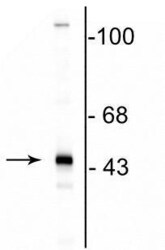
- Experimental details
- Western blot of CNPase in rat brain lysate showing the specific immunolabeling of a band at ~46 kDa corresponding to CNPase polyclonal antibody (Product # PA1-4712).

- Submitted by
- Invitrogen Antibodies (provider)
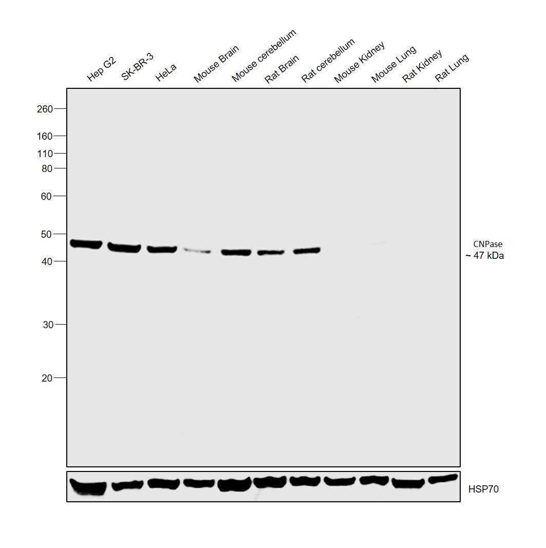
- Main image

- Experimental details
- Western blot was performed using Anti-CNPase Polyclonal Antibody (Product # PA1-4712) and a 47 kDa band corresponding to CNPase was observed in Mouse brain, Mouse cerebellum, Rat Brain and Rat Cerebellum when compared to Mouse Kidney, Mouse Lung, Rat Kidney and Rat Lung which are reported to be low. CNPase is expressed exclusively by oligodendrocytes in the central nervous system. Membrane enriched extracts (30 µg lysate) of Hep G2 (Lane 1), SK-BR-3 (Lane 2), HeLa (Lane 3), tissue extracts of Mouse Brain (Lane 4), Mouse Cerebellum (Lane 5), Rat Brain (Lane 6), Rat Cerebellum (Lane 7), Mouse Kidney (Lane 8), Mouse Lung (Lane 9), Rat Kidney (Lane 10) and Rat Lung (Lane 11) were electrophoresed using Novex® NuPAGE® 4-12 % Bis-Tris gel (Product # NP0322BOX). Resolved proteins were then transferred onto a nitrocellulose membrane (Product # IB23001) by iBlot® 2 Dry Blotting System (Product # IB21001). The blot was probed with the primary antibody (1:1000 dilution) and detected by chemiluminescence with Goat anti-Rabbit IgG (H+L) Superclonal™ Recombinant Secondary Antibody, HRP (Product # A27036, 1:4000 dilution) using the iBright FL 1000 (Product # A32752). Chemiluminescent detection was performed using Novex® ECL Chemiluminescent Substrate Reagent Kit (Product # WP20005).
Supportive validation
- Submitted by
- Invitrogen Antibodies (provider)
- Main image

- Experimental details
- Immunofluorescence analysis of CNPase was performed using 70% confluent log phase Hep G2 cells. The cells were fixed with 4% paraformaldehyde for 10 minutes, permeabilized with 0.1% Triton™ X-100 for 15 minutes, and blocked with 2% BSA for 1 hour at room temperature. The cells were labeled with CNPase Rabbit Polyclonal Antibody (Product # PA1-4712) at 1:100 dilution in 0.1% BSA, incubated at 4 degree Celsius overnight and then with Goat anti-Rabbit IgG (H+L) Highly Cross-Adsorbed Secondary Antibody, Alexa Fluor Plus 488 (Product # A32731) at a dilution of 1:2000 for 45 minutes at room temperature (Panel a: green). Nuclei (Panel b: blue) were stained with ProLong™ Diamond Antifade Mountant with DAPI (Product # P36962). F-actin (Panel c: red) was stained with Rhodamine Phalloidin (Product # R415, 1:300). Panel d represents the merged image showing cytoplasmic localization. Panel e represents control cells with no primary antibody to assess background. The images were captured at 60X magnification.
