Explore
 Validate
 Learn
Learn Western blot
 Immunohistochemistry
Antibody data
- Antibody Data
- Antigen structure
- References [2]
- Comments [0]
- Validations
- Western blot [1]
- Immunohistochemistry [1]
Submit
Validation data
Reference
Comment
Report error
- Product number
- AMAb91068 - Provider product page

- Provider
- Atlas Antibodies
- Proper citation
- Atlas Antibodies Cat#AMAb91068, RRID:AB_2665787
- Product name
- Anti-CNP
- Antibody type
- Monoclonal
- Description
- Monoclonal Antibody against Human CNP, Clone ID: CL2871, Gene description: 2',3'-cyclic nucleotide 3' phosphodiesterase, Validated applications: IHC, WB, Uniprot ID: P09543, Storage: Store at +4°C for short term storage. Long time storage is recommended at -20°C.
- Reactivity
- Human, Mouse, Rat
- Host
- Mouse
- Conjugate
- Unconjugated
- Isotype
- IgG
- Antibody clone number
- CL2871
- Vial size
- 100 µl
- Concentration
- 1.0 mg/ml
- Storage
- Store at +4°C for short term storage. Long time storage is recommended at -20°C.
- Handling
- The antibody solution should be gently mixed before use.
Submitted references Myelin basic protein mRNA levels affect myelin sheath dimensions, architecture, plasticity, and density of resident glial cells
Activin receptors regulate the oligodendrocyte lineage in health and disease.
Bagheri H, Friedman H, Hadwen A, Jarweh C, Cooper E, Oprea L, Guerrier C, Khadra A, Collin A, Cohen‐Adad J, Young A, Victoriano G, Swire M, Jarjour A, Bechler M, Pryce R, Chaurand P, Cougnaud L, Vuckovic D, Wilion E, Greene O, Nishiyama A, Benmamar‐Badel A, Owens T, Grouza V, Tuznik M, Liu H, Rudko D, Zhang J, Siminovitch K, Peterson A
Glia 2024;72(10):1893-1914
Glia 2024;72(10):1893-1914
Activin receptors regulate the oligodendrocyte lineage in health and disease.
Dillenburg A, Ireland G, Holloway RK, Davies CL, Evans FL, Swire M, Bechler ME, Soong D, Yuen TJ, Su GH, Becher JC, Smith C, Williams A, Miron VE
Acta neuropathologica 2018 Jun;135(6):887-906
Acta neuropathologica 2018 Jun;135(6):887-906
No comments: Submit comment
Enhanced validation
- Submitted by
- Atlas Antibodies (provider)
- Enhanced method
- Genetic validation
- Main image
- Experimental details
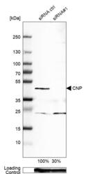
- Western blot analysis in A-549 cells transfected with control siRNA, target specific siRNA probe #1, using Anti-CNP antibody. Remaining relative intensity is presented. Loading control: Anti-GAPDH.
- Sample type
- Human
- Protocol
- Protocol

Supportive validation
- Submitted by
- Atlas Antibodies (provider)
- Enhanced method
- Orthogonal validation
- Main image

- Experimental details
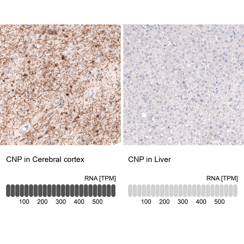
- Immunohistochemistry analysis in human cerebral cortex and liver tissues using AMAb91068 antibody. Corresponding CNP RNA-seq data are presented for the same tissues.
- Sample type
- Human
- Protocol
- Protocol