 Explore
 Validate
 Learn
Learn Western blot
Antibody data
- Antibody Data
- Antigen structure
- References [0]
- Comments [0]
- Validations
- Western blot [5]
- Immunocytochemistry [1]
- Immunohistochemistry [3]
Submit
Validation data
Reference
Comment
Report error
- Product number
- PA5-27972 - Provider product page

- Provider
- Invitrogen Antibodies
- Product name
- CNPase Polyclonal Antibody
- Antibody type
- Polyclonal
- Antigen
- Recombinant protein fragment
- Description
- Recommended positive controls: HeLa, Molt-4, mouse brain, rat brain. Predicted reactivity: Mouse (82%), Rat (84%), Rhesus Monkey (97%), Bovine (89%). Store product as a concentrated solution. Centrifuge briefly prior to opening the vial.
- Reactivity
- Human, Mouse, Rat
- Host
- Rabbit
- Isotype
- IgG
- Vial size
- 100 µL
- Concentration
- 0.41 mg/mL
- Storage
- Store at 4°C short term. For long term storage, store at -20°C, avoiding freeze/thaw cycles.
No comments: Submit comment
Supportive validation
- Submitted by
- Invitrogen Antibodies (provider)
- Main image

- Experimental details
- Western blot analysis of CNP using 20 µg of mouse brain lysate. Samples were loaded onto a 10% SDS-PAGE gel and probed with a CNP polyclonal antibody (Product # PA5-27972) at a dilution of 1:5000.

- Submitted by
- Invitrogen Antibodies (provider)
- Main image

- Experimental details
- Western blot analysis of CNP using 30 µg of MOLT4 lysate. Samples were loaded onto a 10% SDS-PAGE gel and probed with a CNP polyclonal antibody (Product # PA5-27972) at a dilution of 1:1000.

- Submitted by
- Invitrogen Antibodies (provider)
- Main image

- Experimental details
- Western Blot analysis of CNPase was performed by separating 50 µg of various tissue extracts by 10% SDS-PAGE. Proteins were transferred to a membrane and probed with a CNPase Polyclonal Antibody (Product # PA5-27972) at a dilution of 1:5,000.

- Submitted by
- Invitrogen Antibodies (provider)
- Main image

- Experimental details
- Western Blot using CNPase Polyclonal Antibody (Product # PA5-27972). Various whole cell extracts (30 µg) were separated by 10% SDS-PAGE, and the membrane was blotted with CNPase Polyclonal Antibody (Product # PA5-27972) diluted at 1:1,000. The HRP-conjugated anti-rabbit IgG antibody was used to detect the primary antibody.

- Submitted by
- Invitrogen Antibodies (provider)
- Main image

- Experimental details
- Western blot was performed using Anti-CNPase Polyclonal Antibody (Product # PA5-27972) and a 47 kDa band corresponding to CNPase was observed in Mouse brain and Mouse cerebellum when compared to Mouse Kidney, Mouse Lung and Mouse Colon which are reported to be low. CNPase is expressed exclusively by oligodendrocytes in the central nervous system. Membrane enriched extracts (30 µg lysate) of MOLT-4 (Lane 1), Hep G2 (Lane 2), SK-BR-3 (Lane 3), HeLa (Lane 4), tissue extracts of Mouse Brain (Lane 5), Mouse Cerebellum (Lane 6), Mouse Kidney (Lane 7), Mouse Lung (Lane 8) and Mouse Colon (Lane 9) were electrophoresed using Novex® NuPAGE® 4-12 % Bis-Tris gel (Product # NP0322BOX). Resolved proteins were then transferred onto a nitrocellulose membrane (Product # IB23001) by iBlot® 2 Dry Blotting System (Product # IB21001). The blot was probed with the primary antibody (1:5000 dilution) and detected by chemiluminescence with Goat anti-Rabbit IgG (H+L) Superclonal™ Recombinant Secondary Antibody, HRP (Product # A27036, 1:4000 dilution) using the iBright FL 1000 (Product # A32752). Chemiluminescent detection was performed using Novex® ECL Chemiluminescent Substrate Reagent Kit (Product # WP20005).

Supportive validation
- Submitted by
- Invitrogen Antibodies (provider)
- Main image

- Experimental details
- Immunofluorescence analysis of CNPase was performed using 70% confluent log phase Hep G2 cells. The cells were fixed with 4% paraformaldehyde for 10 minutes, permeabilized with 0.1% Triton™ X-100 for 15 minutes, and blocked with 2% BSA for 1 hour at room temperature. The cells were labeled with CNPase Rabbit Polyclonal Antibody (Product # PA5-27972) at 5 µg/mL in 0.1% BSA, incubated at 4 degree Celsius overnight and then with Goat anti-Rabbit IgG (H+L) Highly Cross-Adsorbed Secondary Antibody, Alexa Fluor Plus 488 (Product # A32731) at a dilution of 1:2000 for 45 minutes at room temperature (Panel a: green). Nuclei (Panel b: blue) were stained with ProLong™ Diamond Antifade Mountant with DAPI (Product # P36962). F-actin (Panel c: red) was stained with Rhodamine Phalloidin (Product # R415, 1:300). Panel d represents the merged image showing cytoplasmic localization. Panel e represents control cells with no primary antibody to assess background. The images were captured at 60X magnification.

Supportive validation
- Submitted by
- Invitrogen Antibodies (provider)
- Main image

- Experimental details
- Immunohistochemistry (Frozen) analysis of CNPase was performed in frozen-sectioned adult mouse cerebellum tissue using CNPase Polyclonal Antibody (Product # PA5-27972) at a dilution of 1:250 (Green). Red: beta Tubulin 3/ TUJ1, stained by beta Tubulin 3/ TUJ1 antibody diluted at 1:500. Blue: Fluoroshield with DAPI.

- Submitted by
- Invitrogen Antibodies (provider)
- Main image

- Experimental details
- Immunohistochemistry (Frozen) analysis of CNPase was performed in frozen-sectioned adult mouse hippocampus tissue using CNPase Polyclonal Antibody (Product # PA5-27972) at a dilution of 1:250 (Green). Red: NeuN, stained by NeuN antibody diluted at 1:500.

- Submitted by
- Invitrogen Antibodies (provider)
- Main image
- Experimental details
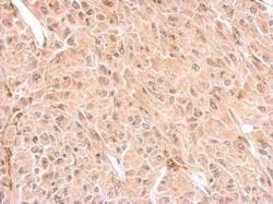
- CNP antibody detects CNP protein at cytosol on U87 xenograft by immunohistochemical analysis. Sample: Paraffin-embedded U87 xenograft. CNP antibody (Product # PA5-27972) dilution: 1:500. Antigen Retrieval: EDTA based buffer, pH 8.0, 15 min.
