Explore
 Validate
 Learn
Learn Western blot
 Immunocytochemistry
Antibody data
- Antibody Data
- Antigen structure
- References [3]
- Comments [0]
- Validations
- Immunocytochemistry [4]
- Immunohistochemistry [1]
- Flow cytometry [3]
- Other assay [2]
Submit
Validation data
Reference
Comment
Report error
- Product number
- 700179 - Provider product page

- Provider
- Invitrogen Antibodies
- Product name
- Cullin 2 Recombinant Rabbit Monoclonal Antibody (50H17L12)
- Antibody type
- Monoclonal
- Antigen
- Synthetic peptide
- Description
- This antibody is predicted to react with bovine, canine, chimpanzee, equine, orangutan, primate, Rhesus monkey and Xenopus based on sequence homology. Intact IgG appears on a non-reducing gel as ~150 kDa band and upon reduction generating a ~25 kDa light chain band and a ~50 kDa heavy chain. Recombinant rabbit monoclonal antibodies are produced using in vitro expression systems. The expression systems are developed by cloning in the specific antibody DNA sequences from immunoreactive rabbits. Then, individual clones are screened to select the best candidates for production. The advantages of using recombinant rabbit monoclonal antibodies include: better specificity and sensitivity, lot-to-lot consistency, animal origin-free formulations, and broader immunoreactivity to diverse targets due to larger rabbit immune repertoire.
- Reactivity
- Human, Mouse, Rat
- Host
- Rabbit
- Isotype
- IgG
- Antibody clone number
- 50H17L12
- Vial size
- 100 μg
- Concentration
- 0.5 mg/mL
- Storage
- Store at 4°C short term. For long term storage, store at -20°C, avoiding freeze/thaw cycles.
Submitted references Unanchored tri-NEDD8 inhibits PARP-1 to protect from oxidative stress-induced cell death.
Differential PROTAC substrate specificity dictated by orientation of recruited E3 ligase.
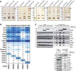
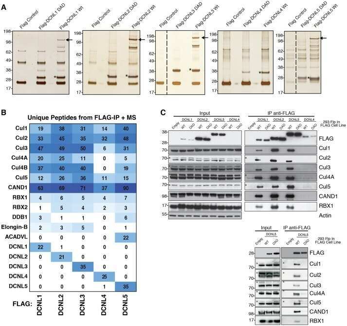
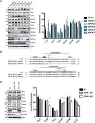
Characterization of the mammalian family of DCN-type NEDD8 E3 ligases.
Keuss MJ, Hjerpe R, Hsia O, Gourlay R, Burchmore R, Trost M, Kurz T
The EMBO journal 2019 Mar 15;38(6)
The EMBO journal 2019 Mar 15;38(6)
Differential PROTAC substrate specificity dictated by orientation of recruited E3 ligase.
Smith BE, Wang SL, Jaime-Figueroa S, Harbin A, Wang J, Hamman BD, Crews CM
Nature communications 2019 Jan 10;10(1):131
Nature communications 2019 Jan 10;10(1):131
Characterization of the mammalian family of DCN-type NEDD8 E3 ligases.
Keuss MJ, Thomas Y, Mcarthur R, Wood NT, Knebel A, Kurz T
Journal of cell science 2016 Apr 1;129(7):1441-54
Journal of cell science 2016 Apr 1;129(7):1441-54
No comments: Submit comment
Supportive validation
- Submitted by
- Invitrogen Antibodies (provider)
- Main image
- Experimental details
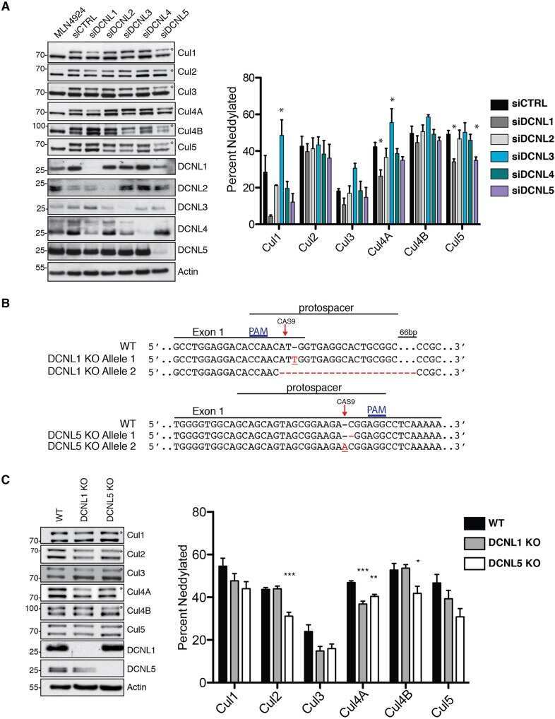
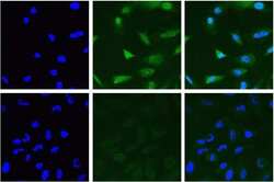
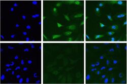
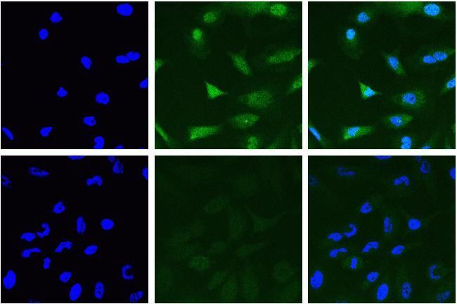
- Immunofluorescent analysis of Cullin-2 in HeLa cells using a Cullin-2 recombinant rabbit monoclonal antibody (Product # 700179) at a dilution of 10 µg/mL in the absence of peptide (top) and presence of immunogenic peptide (bottom), followed by detection using an Alexa Fluor 488-conjugated goat anti-rabbit secondary antibody at a dilution of 1:1000. Actin was stained with Alexa Fluor 568 Phalloidin (Product # A12380). Hoechst only (blue, left), AF488 signal only (green, middle) and composite image with Phalloidin (right).
- Submitted by
- Invitrogen Antibodies (provider)
- Main image
- Experimental details
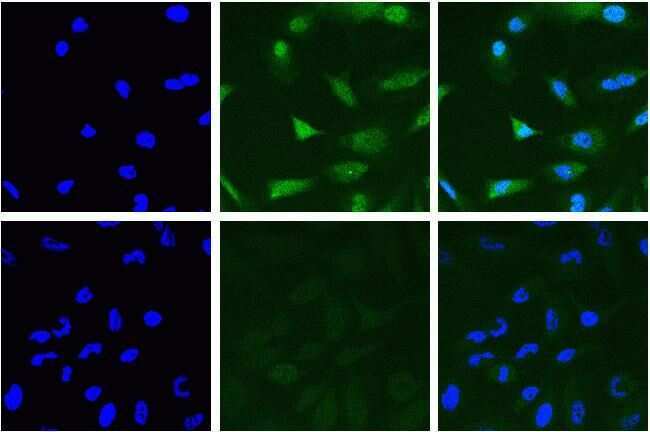
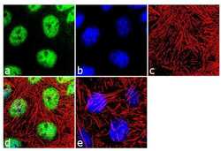
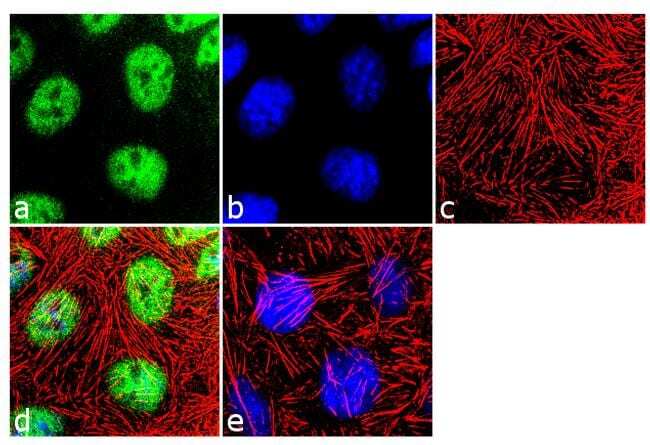
- Immunofluorescence analysis of CUL-2 was performed using 70% confluent log phase Caco-2 cells. The cells were fixed with 4% paraformaldehyde for 10 minutes, permeabilized with 0.1% Triton™ X-100 for 10 minutes, and blocked with 2 % BSA for 1 hour at room temperature. The cells were labeled with CUL-2 (50H17L12) Recombinant Rabbit Monoclonal Antibody (Product # 700179) at 2 µg/mL in 0.1% BSA and incubated for 3 hours at room temperature and then labeled with Goat anti-Rabbit IgG (H+L) Superclonal™ Secondary Antibody, Alexa Fluor® 488 conjugate (Product # A27034) a dilution of 1:2000 for 45 minutes at room temperature (Panel a: green). Nuclei (Panel b: blue) were stained with SlowFade® Gold Antifade Mountant with DAPI (Product # S36938). F-actin (Panel c: red) was stained with Alexa Fluor® 555 Rhodamine Phalloidin (Product # R415, 1:300). Panel d represents the merged image showing nuclear localization. Panel e shows the no primary antibody control. The images were captured at 60X magnification.
- Submitted by
- Invitrogen Antibodies (provider)
- Main image
- Experimental details
- Immunofluorescent analysis of Cullin-2 in HeLa cells using a Cullin-2 recombinant rabbit monoclonal antibody (Product # 700179) at a dilution of 10 µg/mL in the absence of peptide (top) and presence of immunogenic peptide (bottom), followed by detection using an Alexa Fluor 488-conjugated goat anti-rabbit secondary antibody at a dilution of 1:1000. Actin was stained with Alexa Fluor 568 Phalloidin (Product # A12380). Hoechst only (blue, left), AF488 signal only (green, middle) and composite image with Phalloidin (right).
- Submitted by
- Invitrogen Antibodies (provider)
- Main image

- Experimental details
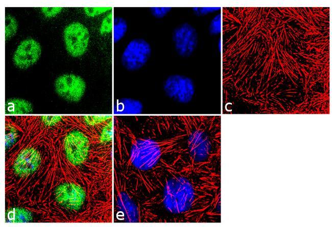
- Immunofluorescence analysis of CUL-2 was performed using 70% confluent log phase Caco-2 cells. The cells were fixed with 4% paraformaldehyde for 10 minutes, permeabilized with 0.1% Triton™ X-100 for 10 minutes, and blocked with 2 % BSA for 1 hour at room temperature. The cells were labeled with CUL-2 (50H17L12) Recombinant Rabbit Monoclonal Antibody (Product # 700179) at 2 µg/mL in 0.1% BSA and incubated for 3 hours at room temperature and then labeled with Goat anti-Rabbit IgG (Heavy Chain) Superclonal™ Secondary Antibody, Alexa Fluor® 488 conjugate (Product # A27034) a dilution of 1:2000 for 45 minutes at room temperature (Panel a: green). Nuclei (Panel b: blue) were stained with SlowFade® Gold Antifade Mountant with DAPI (Product # S36938). F-actin (Panel c: red) was stained with Alexa Fluor® 555 Rhodamine Phalloidin (Product # R415, 1:300). Panel d represents the merged image showing nuclear localization. Panel e shows the no primary antibody control. The images were captured at 60X magnification.
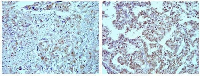
Supportive validation
- Submitted by
- Invitrogen Antibodies (provider)
- Main image
- Experimental details
- Immunohistochemistry analysis of Cullin-2 in formalin-fixed, paraffin-embedded human lung (left) and ovarian carcimona (right) using a Cullin-2 monoclonal antibody (Product # 700179) at a dilution of 5 µg/mL. Tissues were pretreated with EDTA and staining was visualized using DAB. Images were taken at a magnification of 20x. Results show strong nuclear staining in tumor cells.
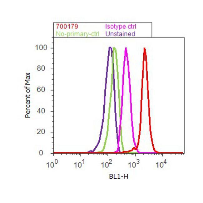
Supportive validation
- Submitted by
- Invitrogen Antibodies (provider)
- Main image

- Experimental details
- Flow cytometry analysis of Cul-2 was done on K562 cells. Cells were fixed with 70% ethanol for 10 minutes, permeabilized with 0.25% Triton™ X-100 for 20 minutes, and blocked with 5% BSA for 30 minutes at room temperature. Cells were labeled with ABfinity™ Cul-2 Recombinant Rabbit Monoclonal Antibody (700179, red histogram) or with rabbit isotype control (pink histogram) at 3-5 ug/million cells in 2.5% BSA. After incubation at room temperature for 2 hours, the cells were labeled with Alexa Fluor® 488 Goat Anti-Rabbit Secondary Antibody (A11008) at a dilution of 1:400 for 30 minutes at room temperature. The representative 10,000 cells were acquired and analyzed for each sample using an Attune® Acoustic Focusing Cytometer. The purple histogram represents unstained control cells and the green histogram represents no-primary-antibody control.
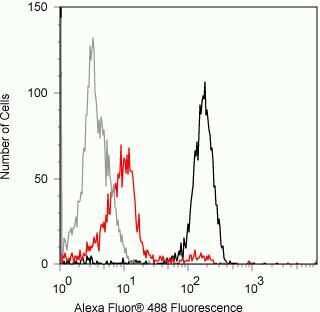
- Submitted by
- Invitrogen Antibodies (provider)
- Main image

- Experimental details
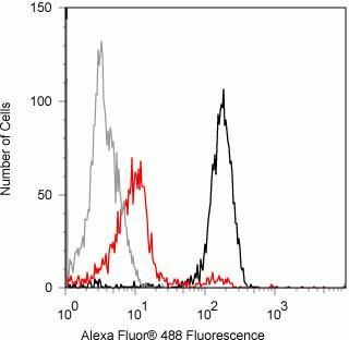
- Flow cytometry analysis of Cullin-2 in Jurkat cells using a Cullin-2 recombinant rabbit monoclonal antibody (Product # 700179) at a dilution of 0.5 µg. Cells were fixed and permeabilized using FIX & PERM (Product # GAS-004) reagent, and detection was performed using an Alexa Fluor 488 goat anti-rabbit IgG (black) compared to a control without primary antibody (gray). Pre-incubation with the immunogenic peptide decreased the signal (red).
- Submitted by
- Invitrogen Antibodies (provider)
- Main image

- Experimental details
- Flow cytometry analysis of Cullin-2 in Jurkat cells using a Cullin-2 recombinant rabbit monoclonal antibody (Product # 700179) at a dilution of 0.5 µg. Cells were fixed and permeabilized using FIX & PERM (Product # GAS-004) reagent, and detection was performed using an Alexa Fluor 488 goat anti-rabbit IgG (black) compared to a control without primary antibody (gray). Pre-incubation with the immunogenic peptide decreased the signal (red).
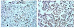
Supportive validation
- Submitted by
- Invitrogen Antibodies (provider)
- Main image
- Experimental details
- NULL
- Submitted by
- Invitrogen Antibodies (provider)
- Main image
- Experimental details
- NULL