Explore
 Validate
 Learn
Learn Western blot
Antibody data
- Antibody Data
- Antigen structure
- References [0]
- Comments [0]
- Validations
- Western blot [1]
- Immunohistochemistry [1]
Submit
Validation data
Reference
Comment
Report error
- Product number
- AF5528 - Provider product page

- Provider
- R&D Systems
- Product name
- Human beta-Synuclein Antibody
- Antibody type
- Polyclonal
- Description
- Antigen Affinity-purified. Detects human beta-Synuclein in direct ELISAs and Western blots. In direct ELISAs, approximately 10% cross-reactivity with recombinant human (rh) alpha-Synuclein and less than 1% cross-reactivity with rh gamma-Synuclein is observed.
- Reactivity
- Human
- Host
- Sheep
- Conjugate
- Unconjugated
- Antigen sequence
Q16143- Isotype
- IgG
- Vial size
- 100 ug
- Concentration
- LYOPH
- Storage
- Use a manual defrost freezer and avoid repeated freeze-thaw cycles. 12 months from date of receipt, -20 to -70 °C as supplied. 1 month, 2 to 8 °C under sterile conditions after reconstitution. 6 months, -20 to -70 °C under sterile conditions after reconstitution.
No comments: Submit comment
Supportive validation
- Submitted by
- R&D Systems (provider)
- Main image
- Experimental details
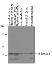
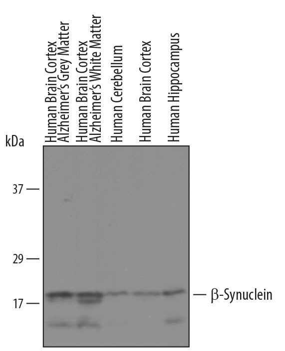
- Detection of Human beta-Synuclein by Western Blot. Western blot shows lysates of human brain cortex, Alzheimer grey matter and white matter, and human cerebellum, brain cortex, and hippocampus tissue. PVDF membrane was probed with 1 µg/mL of Human beta-Synuclein Antigen Affinity-purified Polyclonal Antibody (Catalog # AF5528) followed by HRP-conjugated Anti-Sheep IgG Secondary Antibody (Catalog # HAF016). A specific band was detected for beta-Synuclein at approximately 19 kDa (as indicated). This experiment was conducted under reducing conditions and using Immunoblot Buffer Group 8.
Supportive validation
- Submitted by
- R&D Systems (provider)
- Main image

- Experimental details
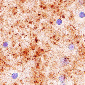
- beta-Synuclein in Human Brain. beta-Synuclein was detected in immersion fixed paraffin-embedded sections of human brain (globus pallidus) using Sheep Anti-Human beta-Synuclein Antigen Affinity-purified Polyclonal Antibody (Catalog # AF5528) at 1.7 µg/mL overnight at 4 °C. Tissue was stained using the Anti-Sheep HRP-DAB Cell & Tissue Staining Kit (brown; Catalog # CTS019) and counterstained with hematoxylin (blue). Specific labeling was localized to the presynaptic terminals. View our protocol for Chromogenic IHC Staining of Paraffin-embedded Tissue Sections.