Explore
 Validate
 Learn
Learn Western blot
 ELISA
ELISA Immunocytochemistry
Antibody data
- Antibody Data
- Antigen structure
- References [13]
- Comments [0]
- Validations
- Immunocytochemistry [2]
- Other assay [8]
Submit
Validation data
Reference
Comment
Report error
- Product number
- 71-9000 - Provider product page

- Provider
- Invitrogen Antibodies
- Product name
- FHIT Polyclonal Antibody
- Antibody type
- Polyclonal
- Antigen
- Other
- Reactivity
- Human, Rat
- Host
- Rabbit
- Isotype
- IgG
- Vial size
- 100 μg
- Concentration
- 0.25 mg/mL
- Storage
- -20°C
Submitted references Fhit induces the reciprocal suppressions between Lin28/Let-7 and miR-17/92miR.
Restoration of MHC-I on Tumor Cells by Fhit Transfection Promotes Immune Rejection and Acts as an Individualized Immunotherapeutic Vaccine.
Upregulation of miR-130b Contributes to Risk of Poor Prognosis and Racial Disparity in African-American Prostate Cancer.
Fhit regulates EMT targets through an EGFR/Src/ERK/Slug signaling axis in human bronchial cells.
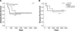
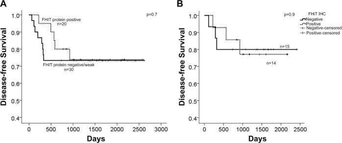
Reduced FHIT expression is associated with mismatch repair deficient and high CpG island methylator phenotype colorectal cancer.
Differential protein immunoexpression profiles in appendiceal mucinous neoplasms: a special reference to classification and predictive factors.
Increased sensitivity to cisplatin in non-small cell lung cancer cell lines after FHIT gene transfer.
Protein expression profiling identifies subclasses of breast cancer and predicts prognosis.
The apoptotic pathway triggered by the Fhit protein in lung cancer cell lines is not affected by Bcl-2 or Bcl-x(L) overexpression.
Synergistic tumor suppression by coexpression of FHIT and p53 coincides with FHIT-mediated MDM2 inactivation and p53 stabilization in human non-small cell lung cancer cells.
Loss of fragile histidine triad protein in human hepatocellular carcinoma.
Restoration of fragile histidine triad (FHIT) expression induces apoptosis and suppresses tumorigenicity in lung and cervical cancer cell lines.
Alterations of the fragile histidine triad gene, FHIT, and its encoded products contribute to testicular germ cell tumorigenesis.
Chae HJ, Seo JB, Kim SH, Jeon YJ, Suh SS
International journal of medical sciences 2021;18(3):706-714
International journal of medical sciences 2021;18(3):706-714
Restoration of MHC-I on Tumor Cells by Fhit Transfection Promotes Immune Rejection and Acts as an Individualized Immunotherapeutic Vaccine.
Pulido M, Chamorro V, Romero I, Algarra I, S-Montalvo A, Collado A, Garrido F, Garcia-Lora AM
Cancers 2020 Jun 12;12(6)
Cancers 2020 Jun 12;12(6)
Upregulation of miR-130b Contributes to Risk of Poor Prognosis and Racial Disparity in African-American Prostate Cancer.
Hashimoto Y, Shiina M, Dasgupta P, Kulkarni P, Kato T, Wong RK, Tanaka Y, Shahryari V, Maekawa S, Yamamura S, Saini S, Deng G, Tabatabai ZL, Majid S, Dahiya R
Cancer prevention research (Philadelphia, Pa.) 2019 Sep;12(9):585-598
Cancer prevention research (Philadelphia, Pa.) 2019 Sep;12(9):585-598
Fhit regulates EMT targets through an EGFR/Src/ERK/Slug signaling axis in human bronchial cells.
Joannes A, Grelet S, Duca L, Gilles C, Kileztky C, Dalstein V, Birembaut P, Polette M, Nawrocki-Raby B
Molecular cancer research : MCR 2014 May;12(5):775-83
Molecular cancer research : MCR 2014 May;12(5):775-83
Reduced FHIT expression is associated with mismatch repair deficient and high CpG island methylator phenotype colorectal cancer.
Al-Temaimi RA, Jacob S, Al-Ali W, Thomas DA, Al-Mulla F
The journal of histochemistry and cytochemistry : official journal of the Histochemistry Society 2013 Sep;61(9):627-38
The journal of histochemistry and cytochemistry : official journal of the Histochemistry Society 2013 Sep;61(9):627-38
Differential protein immunoexpression profiles in appendiceal mucinous neoplasms: a special reference to classification and predictive factors.
Yoon SO, Kim BH, Lee HS, Kang GH, Kim WH, Kim YA, Kim JE, Chang MS
Modern pathology : an official journal of the United States and Canadian Academy of Pathology, Inc 2009 Aug;22(8):1102-12
Modern pathology : an official journal of the United States and Canadian Academy of Pathology, Inc 2009 Aug;22(8):1102-12
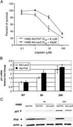
Increased sensitivity to cisplatin in non-small cell lung cancer cell lines after FHIT gene transfer.
Andriani F, Perego P, Carenini N, Sozzi G, Roz L
Neoplasia (New York, N.Y.) 2006 Jan;8(1):9-17
Neoplasia (New York, N.Y.) 2006 Jan;8(1):9-17
Protein expression profiling identifies subclasses of breast cancer and predicts prognosis.
Jacquemier J, Ginestier C, Rougemont J, Bardou VJ, Charafe-Jauffret E, Geneix J, Adélaïde J, Koki A, Houvenaeghel G, Hassoun J, Maraninchi D, Viens P, Birnbaum D, Bertucci F
Cancer research 2005 Feb 1;65(3):767-79
Cancer research 2005 Feb 1;65(3):767-79
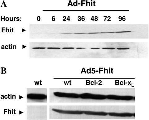
The apoptotic pathway triggered by the Fhit protein in lung cancer cell lines is not affected by Bcl-2 or Bcl-x(L) overexpression.
Roz L, Andriani F, Ferreira CG, Giaccone G, Sozzi G
Oncogene 2004 Dec 2;23(56):9102-10
Oncogene 2004 Dec 2;23(56):9102-10
Synergistic tumor suppression by coexpression of FHIT and p53 coincides with FHIT-mediated MDM2 inactivation and p53 stabilization in human non-small cell lung cancer cells.
Nishizaki M, Sasaki J, Fang B, Atkinson EN, Minna JD, Roth JA, Ji L
Cancer research 2004 Aug 15;64(16):5745-52
Cancer research 2004 Aug 15;64(16):5745-52
Loss of fragile histidine triad protein in human hepatocellular carcinoma.
Zhao P, Song X, Nin YY, Lu YL, Li XH
World journal of gastroenterology 2003 Jun;9(6):1216-9
World journal of gastroenterology 2003 Jun;9(6):1216-9
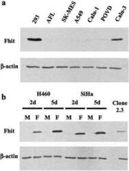
Restoration of fragile histidine triad (FHIT) expression induces apoptosis and suppresses tumorigenicity in lung and cervical cancer cell lines.
Roz L, Gramegna M, Ishii H, Croce CM, Sozzi G
Proceedings of the National Academy of Sciences of the United States of America 2002 Mar 19;99(6):3615-20
Proceedings of the National Academy of Sciences of the United States of America 2002 Mar 19;99(6):3615-20
Alterations of the fragile histidine triad gene, FHIT, and its encoded products contribute to testicular germ cell tumorigenesis.
Kraggerud SM, Aman P, Holm R, Stenwig AE, Fosså SD, Nesland JM, Lothe RA
Cancer research 2002 Jan 15;62(2):512-7
Cancer research 2002 Jan 15;62(2):512-7
No comments: Submit comment
Supportive validation
- Submitted by
- Invitrogen Antibodies (provider)
- Main image
- Experimental details
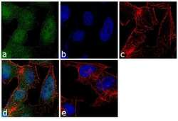
- Immunofluorescence analysis of FHIT Polyclonal Antibody was performed using 70% confluent log phase Hep G2 cells. The cells were fixed with 4% paraformaldehyde for 10 minutes, permeabilized with 0.1% Triton X-100 for 10 minutes, and blocked with 1% BSA for 1 hour at room temperature. The cells were labeled with FHIT Rabbit Polyclonal Antibody at 2 µg/mL in 0.1% BSA and incubated for 3 hours at room temperature and then labeled with Goat anti-Rabbit IgG (H+L) Superclonal Secondary Antibody, Alexa Fluor® 488 conjugate (Product # A27034) at a dilution of 1:2000 for 45 minutes at room temperature (Panel a: green). Nuclei (Panel b: blue) were stained with SlowFade® Gold Antifade Mountant with DAPI (Product # S36938). F-actin (Panel c: red) was stained with Rhodamine Phalloidin (Product # R415, 1:300). Panel d represents the merged image showing nuclear and cytoplasmic localization. Panel e shows the no primary antibody control. The images were captured at 60X magnification.
- Submitted by
- Invitrogen Antibodies (provider)
- Main image
- Experimental details
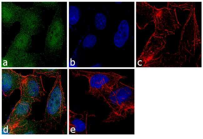
- Immunofluorescence analysis of FHIT Polyclonal Antibody was performed using 70% confluent log phase Hep G2 cells. The cells were fixed with 4% paraformaldehyde for 10 minutes, permeabilized with 0.1% Triton X-100 for 10 minutes, and blocked with 1% BSA for 1 hour at room temperature. The cells were labeled with FHIT Rabbit Polyclonal Antibody at 2 µg/mL in 0.1% BSA and incubated for 3 hours at room temperature and then labeled with Goat anti-Rabbit IgG (Heavy Chain) Superclonal Secondary Antibody, Alexa Fluor® 488 conjugate (Product # A27034) at a dilution of 1:2000 for 45 minutes at room temperature (Panel a: green). Nuclei (Panel b: blue) were stained with SlowFade® Gold Antifade Mountant with DAPI (Product # S36938). F-actin (Panel c: red) was stained with Rhodamine Phalloidin (Product # R415, 1:300). Panel d represents the merged image showing nuclear and cytoplasmic localization. Panel e shows the no primary antibody control. The images were captured at 60X magnification.

Supportive validation
- Submitted by
- Invitrogen Antibodies (provider)
- Main image
- Experimental details
- NULL
- Submitted by
- Invitrogen Antibodies (provider)
- Main image
- Experimental details
- NULL

- Submitted by
- Invitrogen Antibodies (provider)
- Main image

- Experimental details
- NULL
- Submitted by
- Invitrogen Antibodies (provider)
- Main image
- Experimental details
- NULL
- Submitted by
- Invitrogen Antibodies (provider)
- Main image

- Experimental details
- NULL
- Submitted by
- Invitrogen Antibodies (provider)
- Main image

- Experimental details
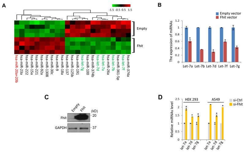
- Figure 1 Suppressed let-7 miRNAs in response to Fhit overexpression. (A) Unsupervised clustering of miRNAs in HCT116 expressing Fhit. HCT116-Fhit stable cells were generated by transduction of lenti-Fhit virus followed by puromycin selection for 5 days (A). Subsequently, the total RNAs were extracted and analyzed by miRNA microarray analysis. b and c, Validation of let-7 miRNA suppression by Fhit gene in HCT116 cell line. The cells were transiently transfected by Fhit plasmid for 24 hrs. Subsequently the cells were harvested and prepared for qRT-PCR analysis. (B). In parallel the Fhit expression was validated by Western blot analysis using anti-Fhit antibody (C). (D) The suppression of let-7 family by Fhit deficiency in A549 lung cancer cell line enforced by siFhit siRNA treatment. Three independent experiments were performed in triplicate (n=3). Bars mean +-S.D. and the p-value were obtained by student t-test (*p
- Submitted by
- Invitrogen Antibodies (provider)
- Main image
- Experimental details
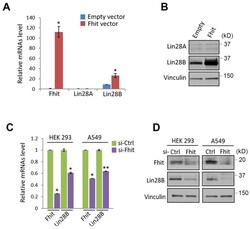
- Figure 2 Fhit-dependent induction of Lin28 protein. (A) Increased Lin28B mRNA in A549 expressing Fhit gene. The cell was transiently transfected by pcDNA3-Fhit plasmid, and Lin28 mRNA expression was analyzed by qRT-PCR analysis using the extracted RNA from A549 expressing Fhit. (B) Increased Lin28B protein in HCT116-Fhit stable cell line. The indicated cells and its parental cell were prepared and subject to Western blot analysis using anti-Lin28B antibody. Lin28B expression was further validated by independent biological replicate. (C and D) Negative regulation of Lin28 in Fhit deficient cell lines. The indicated cells were transiently transfected by siFhit siRNAs. After 48 hrs of transfection, the cells were harvested and Lin28 expression was analyzed by qRT-PCR (C) or Western blot analysis (D). Three independent experiments were performed in triplicate (n=3). Bars mean +-S.D. and the p-value were obtained by student t-test (*p

- Submitted by
- Invitrogen Antibodies (provider)
- Main image
- Experimental details
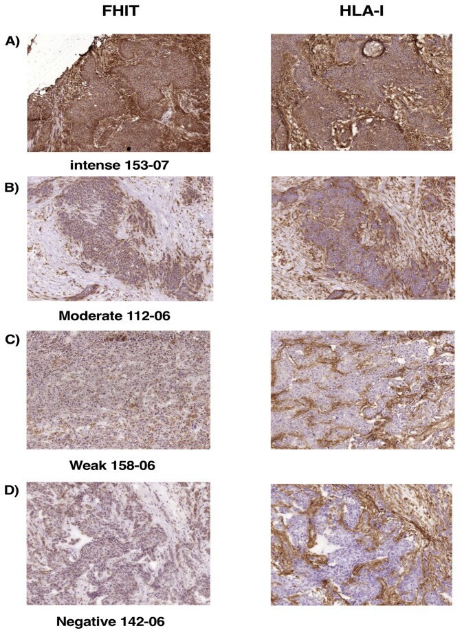
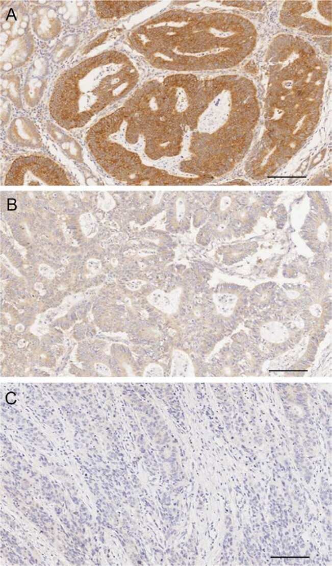
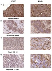
- Figure 6 Expression of FHIT and MHC-I in human breast carcinoma cells analyzed by immunohistochemistry. ( A ) Representative tumor with intense FHIT and HLA-I expressions; ( B ) Representative tumor with moderate FHIT and HLA-I expressions; ( C ) Representative tumor with weak FHIT and HLA-I expressions. ( D ) Representative tumor with negative FHIT and HLA-I expressions. The same tumor cells were analyzed in each case for FHIT and HLA-I expression. Magnification 200x.