 Explore
 Validate
 Learn
Learn Western blot
Antibody data
- Antibody Data
- Antigen structure
- References [3]
- Comments [0]
- Validations
- Western blot [1]
- Immunohistochemistry [6]
Submit
Validation data
Reference
Comment
Report error
- Product number
- NBP1-89062 - Provider product page

- Provider
- Novus Biologicals
- Proper citation
- Novus Cat#NBP1-89062, RRID:AB_11032003
- Product name
- Rabbit Polyclonal FHIT Antibody
- Antibody type
- Polyclonal
- Description
- Immunogen affinity purified. Specificity of human FHIT antibody verified on a Protein Array containing target protein plus 383 other non-specific proteins.
- Reactivity
- Human
- Host
- Rabbit
- Isotype
- IgG
- Vial size
- 0.1 ml
- Storage
- Store at 4C short term. Aliquot and store at -20C long term. Avoid freeze-thaw cycles.
Submitted references High-resolution whole-genome analysis of skull base chordomas implicates FHIT loss in chordoma pathogenesis.
High-resolution whole-genome analysis of skull base chordomas implicates FHIT loss in chordoma pathogenesis.
The protein expression of TRP-1 and galectin-1 in cutaneous malignant melanomas.
Diaz RJ, Guduk M, Romagnuolo R, Smith CA, Northcott P, Shih D, Berisha F, Flanagan A, Munoz DG, Cusimano MD, Pamir MN, Rutka JT
Neoplasia (New York, N.Y.) 2012 Sep;14(9):788-98
Neoplasia (New York, N.Y.) 2012 Sep;14(9):788-98
High-resolution whole-genome analysis of skull base chordomas implicates FHIT loss in chordoma pathogenesis.
Diaz RJ, Guduk M, Romagnuolo R, Smith CA, Northcott P, Shih D, Berisha F, Flanagan A, Munoz DG, Cusimano MD, Pamir MN, Rutka JT
Neoplasia (New York, N.Y.) 2012 Sep;14(9):788-98
Neoplasia (New York, N.Y.) 2012 Sep;14(9):788-98
The protein expression of TRP-1 and galectin-1 in cutaneous malignant melanomas.
Bolander A, Agnarsdóttir M, Strömberg S, Ponten F, Hesselius P, Uhlen M, Bergqvist M
Cancer genomics & proteomics 2008 Nov-Dec;5(6):293-300
Cancer genomics & proteomics 2008 Nov-Dec;5(6):293-300
No comments: Submit comment
Supportive validation
- Submitted by
- Novus Biologicals (provider)
- Main image

- Experimental details
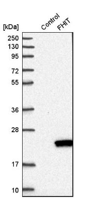
- Western Blot: FHIT Antibody [NBP1-89062] - Analysis in control (vector only transfected HEK293T lysate) and fHIT over-expression lysate (Co-expressed with a C-terminal myc-DDK tag (3.1 kDa) in mammalian HEK293T cells).
Supportive validation
- Submitted by
- Novus Biologicals (provider)
- Main image

- Experimental details
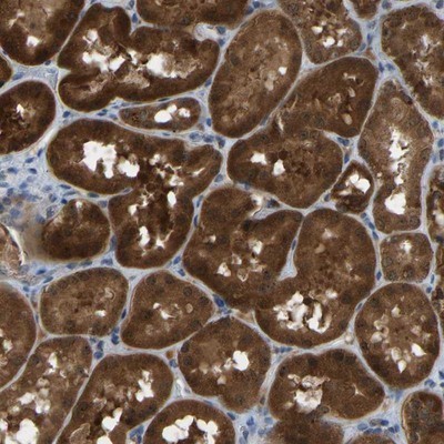
- Immunohistochemistry-Paraffin: FHIT Antibody [NBP1-89062] - Staining of human kidney shows strong cytoplasmic positivity in tubule cells.
- Submitted by
- Novus Biologicals (provider)
- Main image

- Experimental details
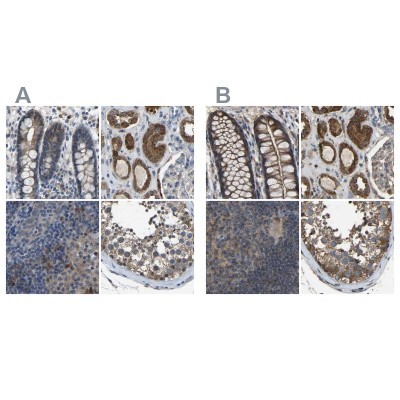
- Immunohistochemistry-Paraffin: FHIT Antibody [NBP1-89062] - Staining of human colon, kidney, lymph node and testis using Anti-FHIT antibody NBP1-89062 (A) shows similar protein distribution across tissues to independent antibody NBP1-89061 (B).
- Submitted by
- Novus Biologicals (provider)
- Main image
- Experimental details
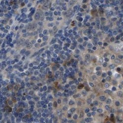
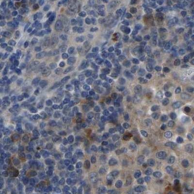
- Immunohistochemistry-Paraffin: FHIT Antibody [NBP1-89062] - Staining of human lymph node.
- Submitted by
- Novus Biologicals (provider)
- Main image
- Experimental details
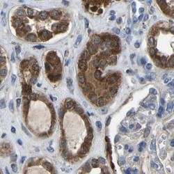
- Immunohistochemistry-Paraffin: FHIT Antibody [NBP1-89062] - Staining of human kidney.

- Submitted by
- Novus Biologicals (provider)
- Main image

- Experimental details
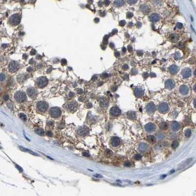
- Immunohistochemistry-Paraffin: FHIT Antibody [NBP1-89062] - Staining of human testis.
- Submitted by
- Novus Biologicals (provider)
- Main image
- Experimental details
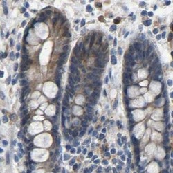
- Immunohistochemistry-Paraffin: FHIT Antibody [NBP1-89062] - Staining of human colon.
