Explore
 Validate
 Learn
Learn Western blot
 ELISA
ELISAAntibody data
- Antibody Data
- Antigen structure
- References [2]
- Comments [0]
- Validations
- Western blot [1]
Submit
Validation data
Reference
Comment
Report error
- Product number
- PB9327 - Provider product page

- Provider
- Boster Biological Technology
- Product name
- Anti-Apolipoprotein E/APOE Antibody Picoband™
- Antibody type
- Polyclonal
- Description
- Polyclonal antibody for Apolipoprotein E/APOE detection. Host: Rabbit.Size: 100μg/vial. Tested applications: WB, IHC-P, IHC-F, FCM, ELISA(Cap). Reactive species: Human. Apolipoprotein E/APOE information: Molecular Weight: 36154 MW; Subcellular Localization: Secreted; Tissue Specificity: Occurs in all lipoprotein fractions in plasma. It constitutes 10-20% of very low density lipoproteins (VLDL) and 1-2% of high density lipoproteins (HDL). APOE is produced in most organs. Significant quantities are produced in liver, brain, spleen, lung, adrenal, ovary, kidney and muscle.
- Reactivity
- Human
- Host
- Rabbit
- Vial size
- 100μg/vial
- Concentration
- Add 0.2ml of distilled water will yield a concentration of 500ug/ml.
- Storage
- At -20°C for one year. After reconstitution, at 4°C for one month. It can also be aliquoted and stored frozen at -20°C for a longer time. Avoid repeated freezing and thawing.
- Handling
- Add 0.2ml of distilled water will yield a concentration of 500ug/ml.
Submitted references Phaseolus vulgaris Erythroagglutinin (PHA-E)-Positive Ceruloplasmin Acts as a Potential Biomarker in Pancreatic Cancer Diagnosis.
Identification of Apolipoprotein E as a Potential Diagnostic Biomarker of Nasopharyngeal Carcinoma.
Sha S, Wang Y, Liu M, Liu G, Fan N, Li Z, Dong W
Cells 2022 Aug 8;11(15)
Cells 2022 Aug 8;11(15)
Identification of Apolipoprotein E as a Potential Diagnostic Biomarker of Nasopharyngeal Carcinoma.
Xue Y, Huang S, Huang J, Li S, Zhang C, Zhou X
Cancer management and research 2020;12:8943-8950
Cancer management and research 2020;12:8943-8950
No comments: Submit comment
Supportive validation
- Submitted by
- Boster Biological Technology (provider)
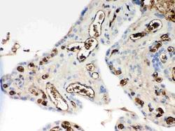
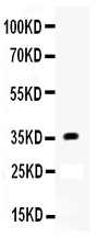
- Main image

- Experimental details
- Western blot analysis of Apolipoprotein E using anti-Apolipoprotein E antibody (PB9327). Electrophoresis was performed on a 5-20% SDS-PAGE gel at 70V (Stacking gel) / 90V (Resolving gel) for 2-3 hours. The sample well of each lane was loaded with 50ug of sample under reducing conditions. Lane 1: Human Placenta Tissue Lysate. After Electrophoresis, proteins were transferred to a Nitrocellulose membrane at 150mA for 50-90 minutes. Blocked the membrane with 5% Non-fat Milk/ TBS for 1.5 hour at RT. The membrane was incubated with rabbit anti-Apolipoprotein E antigen affinity purified polyclonal antibody (Catalog # PB9327) at 0.5 μg/mL overnight at 4°C, then washed with TBS-0.1%Tween 3 times with 5 minutes each and probed with a goat anti-rabbit IgG-HRP secondary antibody at a dilution of 1:10000 for 1.5 hour at RT. The signal is developed using an Enhanced Chemiluminescent detection (ECL) kit (Catalog # EK1002) with Tanon 5200 system. A specific band was detected for Apolipoprotein E at approximately 36KD. The expected band size for Apolipoprotein E is at 36KD.
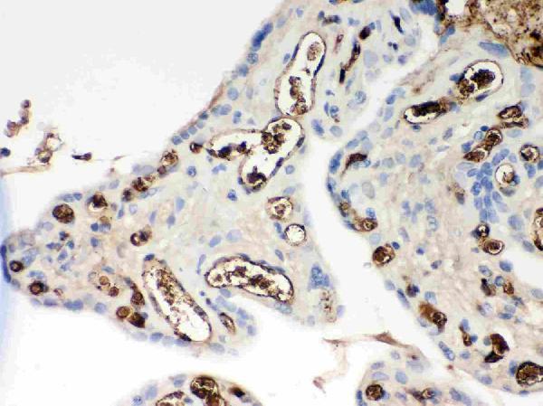
- Additional image