 Explore
 Validate
 Learn
Learn Western blot
 ELISA
ELISAAntibody data
- Antibody Data
- Antigen structure
- References [3]
- Comments [0]
- Validations
- ELISA [5]
- Immunocytochemistry [6]
- Immunoprecipitation [1]
- Immunohistochemistry [1]
- Other assay [2]
Submit
Validation data
Reference
Comment
Report error
- Product number
- PA5-27088 - Provider product page

- Provider
- Invitrogen Antibodies
- Product name
- APOE Polyclonal Antibody
- Antibody type
- Polyclonal
- Antigen
- Synthetic peptide
- Description
- Recommended positive controls: HepG2, Conditional medium from differentiated human primary preadipocyte, human plasma. Store product as a concentrated solution. Centrifuge briefly prior to opening the vial.
- Reactivity
- Human
- Host
- Rabbit
- Isotype
- IgG
- Vial size
- 100 μL
- Concentration
- 0.66 mg/mL
- Storage
- Store at 4°C short term. For long term storage, store at -20°C, avoiding freeze/thaw cycles.
Submitted references Plaque-associated human microglia accumulate lipid droplets in a chimeric model of Alzheimer's disease.
Morphological analysis of Apolipoprotein E binding to Aβ Amyloid using a combination of Surface Plasmon Resonance, Immunogold Labeling and Scanning Electron Microscopy.
Complement-Mediated Regulation of Apolipoprotein E in Cultured Human RPE Cells.
Claes C, Danhash EP, Hasselmann J, Chadarevian JP, Shabestari SK, England WE, Lim TE, Hidalgo JLS, Spitale RC, Davtyan H, Blurton-Jones M
Molecular neurodegeneration 2021 Jul 23;16(1):50
Molecular neurodegeneration 2021 Jul 23;16(1):50
Morphological analysis of Apolipoprotein E binding to Aβ Amyloid using a combination of Surface Plasmon Resonance, Immunogold Labeling and Scanning Electron Microscopy.
Islam T, Gharibyan AL, Lee CC, Olofsson A
BMC biotechnology 2019 Dec 11;19(1):97
BMC biotechnology 2019 Dec 11;19(1):97
Complement-Mediated Regulation of Apolipoprotein E in Cultured Human RPE Cells.
Yang P, Skiba NP, Tewkesbury GM, Treboschi VM, Baciu P, Jaffe GJ
Investigative ophthalmology & visual science 2017 Jun 1;58(7):3073-3085
Investigative ophthalmology & visual science 2017 Jun 1;58(7):3073-3085
No comments: Submit comment
Supportive validation
- Submitted by
- Invitrogen Antibodies (provider)
- Main image

- Experimental details
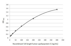
- Sandwich ELISA detection of recombinant full-length Apolipoprotein E protein using APOE Polyclonal Antibody (Product # PA5-27088) as capture antibody at concentration of 5 µg/mL and a mouse monoclonal anti-Apolipoprotein E antibody as detection antibody at concentration of 1 µg/mL. Mouse IgG antibody (HRP) was diluted at 1:10,000 and used to detect the primary antibody.

- Submitted by
- Invitrogen Antibodies (provider)
- Main image

- Experimental details
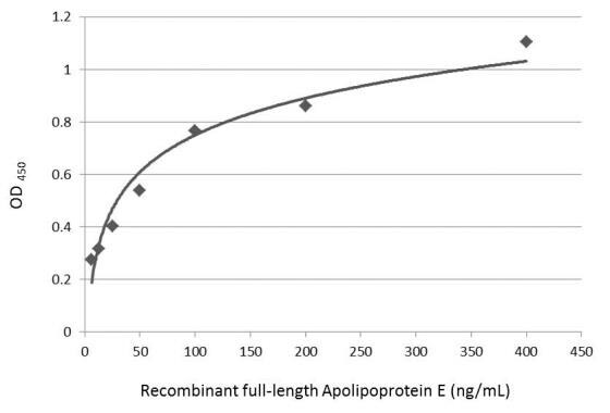
- Sandwich ELISA detection of recombinant full-length human apolipoprotein E using a APOE Monoclonal Antibody as capture antibody at concentration of 5 µg/mL and APOE Polyclonal Antibody (Product # PA5-27088) as detection antibody at concentration of 1 µg/mL. Rabbit IgG antibody (HRP) was diluted at 1:10,000 and used to detect the primary antibody.

- Submitted by
- Invitrogen Antibodies (provider)
- Main image
- Experimental details
- Sandwich ELISA detection of recombinant full-length human apolipoprotein E using APOE Monoclonal Antibody at concentration of 5 µg/mL and APOE Polyclonal Antibody (Product # PA5-27088) as detection antibody at concentration of 1 µg/mL. Rabbit IgG antibody (HRP) was diluted at 1:10,000 and used to detect the primary antibody.

- Submitted by
- Invitrogen Antibodies (provider)
- Main image

- Experimental details
- Sandwich ELISA detection of recombinant full-length Apolipoprotein E protein using APOE Polyclonal Antibody (Product # PA5-27088) as capture antibody at concentration of 5 µg/mL and a mouse monoclonal anti-Apolipoprotein E antibody as detection antibody at concentration of 1 µg/mL. Mouse IgG antibody (HRP) was diluted at 1:10,000 and used to detect the primary antibody.
- Submitted by
- Invitrogen Antibodies (provider)
- Main image

- Experimental details
- Sandwich ELISA detection of recombinant full-length human apolipoprotein E using a APOE Monoclonal Antibody as capture antibody at concentration of 5 µg/mL and APOE Polyclonal Antibody (Product # PA5-27088) as detection antibody at concentration of 1 µg/mL. Rabbit IgG antibody (HRP) was diluted at 1:10,000 and used to detect the primary antibody.

Supportive validation
- Submitted by
- Invitrogen Antibodies (provider)
- Main image
- Experimental details
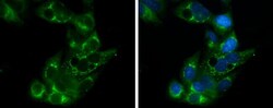
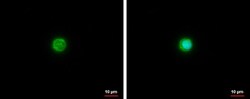
- Immunocytochemistry-Immunofluorescence analysis of APOE was performed in HepG2 cells fixed in 4% paraformaldehyde at RT for 15 min. Green: APOE Polyclonal Antibody (Product # PA5-27088) diluted at 1:500. Blue: Hoechst 33342 staining.
- Submitted by
- Invitrogen Antibodies (provider)
- Main image

- Experimental details
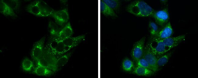
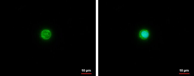
- APOE Polyclonal Antibody [C2C3], C-term detects Apolipoprotein E protein at cytoplasm by immunofluorescent analysis. Sample: THP-1 cells were fixed in ice-cold MeOH for 5 min. Green: Apolipoprotein E protein stained by APOE Polyclonal Antibody [C2C3], C-term (Product # PA5-27088) diluted at 1:500. Blue: Hoechst 33342 staining.

- Submitted by
- Invitrogen Antibodies (provider)
- Main image

- Experimental details
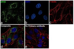
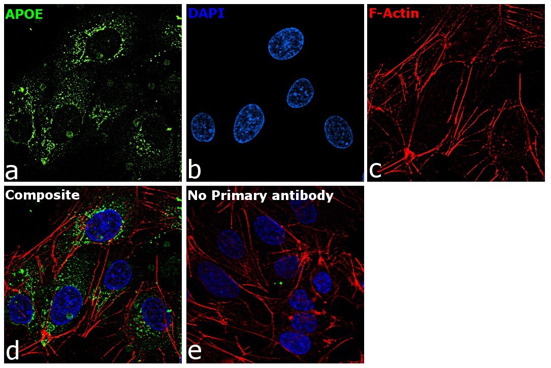
- Immunofluorescence analysis of APOE was performed using 70% confluent log phase Hep G2 cells. The cells were fixed with 4% paraformaldehyde for 10 minutes, permeabilized with 0.1% Triton™ X-100 for 15 minutes, and blocked with 2% BSA for 1 hour at room temperature. The cells were labeled with APOE Rabbit Polyclonal Antibody (Product # PA5-27088) at 5 µg/mL in 0.1% BSA, incubated at 4 degree Celsius overnight and then with Goat anti-Rabbit IgG (H+L) Highly Cross-Adsorbed Secondary Antibody, Alexa Fluor Plus 488 (Product # A32731) at a dilution of 1:2000 for 45 minutes at room temperature (Panel a: green). Nuclei (Panel b: blue) were stained with ProLong™ Diamond Antifade Mountant with DAPI (Product # P36962). F-actin (Panel c: red) was stained with Rhodamine Phalloidin (Product # R415, 1:300). Panel d represents the merged image showing cytoplasmic localization. Panel e represents control cells with no primary antibody to assess background. The images were captured at 60X magnification.

- Submitted by
- Invitrogen Antibodies (provider)
- Main image
- Experimental details
- APOE Polyclonal Antibody [C2C3], C-term detects Apolipoprotein E protein at cytoplasm by immunofluorescent analysis. Sample: THP-1 cells were fixed in ice-cold MeOH for 5 min. Green: Apolipoprotein E protein stained by APOE Polyclonal Antibody [C2C3], C-term (Product # PA5-27088) diluted at 1:500. Blue: Hoechst 33342 staining.
- Submitted by
- Invitrogen Antibodies (provider)
- Main image

- Experimental details
- Immunocytochemistry-Immunofluorescence analysis of APOE was performed in HepG2 cells fixed in 4% paraformaldehyde at RT for 15 min. Green: APOE Polyclonal Antibody (Product # PA5-27088) diluted at 1:500. Blue: Hoechst 33342 staining.

- Submitted by
- Invitrogen Antibodies (provider)
- Main image
- Experimental details
- Immunofluorescence analysis of APOE was performed using 70% confluent log phase Hep G2 cells. The cells were fixed with 4% paraformaldehyde for 10 minutes, permeabilized with 0.1% Triton™ X-100 for 15 minutes, and blocked with 2% BSA for 1 hour at room temperature. The cells were labeled with APOE Rabbit Polyclonal Antibody (Product # PA5-27088) at 5 µg/mL in 0.1% BSA, incubated at 4 degree Celsius overnight and then with Goat anti-Rabbit IgG (H+L) Highly Cross-Adsorbed Secondary Antibody, Alexa Fluor Plus 488 (Product # A32731) at a dilution of 1:2000 for 45 minutes at room temperature (Panel a: green). Nuclei (Panel b: blue) were stained with ProLong™ Diamond Antifade Mountant with DAPI (Product # P36962). F-actin (Panel c: red) was stained with Rhodamine Phalloidin (Product # R415, 1:300). Panel d represents the merged image showing cytoplasmic localization. Panel e represents control cells with no primary antibody to assess background. The images were captured at 60X magnification.
Supportive validation
- Submitted by
- Invitrogen Antibodies (provider)
- Main image

- Experimental details
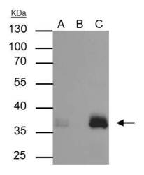
- APOE Polyclonal Antibody immunoprecipitates Apolipoprotein E protein in IP experiments. IP Sample: HepG2 whole cell lysate/extract. A : 30 µg whole cell lysate/extract of Apolipoprotein E protein expressing HepG2 cells. B : Control with 3 µg of pre-immune rabbit IgG. C : Immunoprecipitation of Apolipoprotein E by 3 µg of APOE Polyclonal Antibody (Product # PA5-27088). 10% SDS-PAGE. The immunoprecipitated Apolipoprotein E protein was detected by APOE Polyclonal Antibody (Product # PA5-27088) diluted at 1:1,000. Anti-rabbit IgG (HRP) was used as a secondary reagent.

Supportive validation
- Submitted by
- Invitrogen Antibodies (provider)
- Main image
- Experimental details
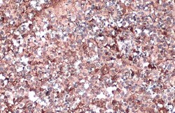
- Immunohistochemistry (Paraffin) analysis of APOE was performed in paraffin-embedded human ovarian cancer tissue using APOE Polyclonal Antibody (Product # PA5-27088) at a dilution of 1:500. Antigen Retrieval: Citrate buffer, pH 6.0, 15 min.

Supportive validation
- Submitted by
- Invitrogen Antibodies (provider)
- Main image
- Experimental details
- APOE Polyclonal Antibody immunoprecipitates Apolipoprotein E protein in IP experiments. IP Sample: HepG2 whole cell lysate/extract. A : 30 µg whole cell lysate/extract of Apolipoprotein E protein expressing HepG2 cells. B : Control with 3 µg of pre-immune rabbit IgG. C : Immunoprecipitation of Apolipoprotein E by 3 µg of APOE Polyclonal Antibody (Product # PA5-27088). 10% SDS-PAGE. The immunoprecipitated Apolipoprotein E protein was detected by APOE Polyclonal Antibody (Product # PA5-27088) diluted at 1:1,000. Anti-rabbit IgG (HRP) was used as a secondary reagent.

- Submitted by
- Invitrogen Antibodies (provider)
- Main image
- Experimental details
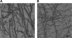
- Fig. 3 SEM analysis of the SPR-chip surface. a Control sample in absence of added ApoE to probe for non-specific binding where the immobilized fibrils on the SPR-chip have been sequentially probed with anti-ApoE antibodies and protein-A conjugated 15 nm gold-beads. b Complete setup where fibrils bound to the SPR-chip have been sequentially probed with ApoE4, anti-ApoE antibodies and protein-A conjugated 15 nm gold-beads. Scale bar is 100 nm
