Explore
 Validate
 Learn
Learn Western blot
 Immunohistochemistry
Antibody data
- Antibody Data
- Antigen structure
- References [2]
- Comments [0]
- Validations
- Western blot [3]
Submit
Validation data
Reference
Comment
Report error
- Product number
- MA1-20377 - Provider product page

- Provider
- Invitrogen Antibodies
- Product name
- Anti-Drebrin Monoclonal Antibody (M2F6)
- Antibody type
- Monoclonal
- Antigen
- Other
- Description
- A suggested positive control for this product is mouse brain tissue extract.
- Reactivity
- Human, Mouse, Rat, Chicken/Avian, Feline, Guinea Pig, Porcine
- Host
- Mouse
- Isotype
- IgG
- Antibody clone number
- M2F6
- Vial size
- 50 µg
- Concentration
- 1 mg/mL
- Storage
- Store at 4°C short term. For long term storage, store at -20°C, avoiding freeze/thaw cycles.
Submitted references The Actin-Binding Protein Drebrin Inhibits Neointimal Hyperplasia.
Selective reduction of drebrin and actin in dendritic spines of hippocampal neurons by activation of 5-HT(2A) receptors.
Stiber JA, Wu JH, Zhang L, Nepliouev I, Zhang ZS, Bryson VG, Brian L, Bentley RC, Gordon-Weeks PR, Rosenberg PB, Freedman NJ
Arteriosclerosis, thrombosis, and vascular biology 2016 May;36(5):984-93
Arteriosclerosis, thrombosis, and vascular biology 2016 May;36(5):984-93
Selective reduction of drebrin and actin in dendritic spines of hippocampal neurons by activation of 5-HT(2A) receptors.
Roppongi RT, Kojima N, Hanamura K, Yamazaki H, Shirao T
Neuroscience letters 2013 Jun 28;547:76-81
Neuroscience letters 2013 Jun 28;547:76-81
No comments: Submit comment
Supportive validation
- Submitted by
- Invitrogen Antibodies (provider)
- Main image

- Experimental details
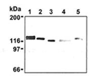
- Western blot analysis of Drebrin in mouse brain (1) NIH/3T3 (2), Jurkat (3), HeLa (4), and PC12 (5) using a Drebrin monoclonal antibody (Product # MA1-20377).
- Submitted by
- Invitrogen Antibodies (provider)
- Main image
- Experimental details
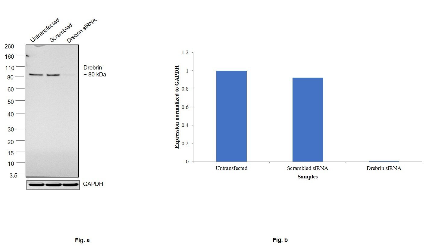
- Knockdown of Anti-Drebrin Monoclonal Antibody (M2F6) was achieved by transfecting HEK-293 cells with Drebrin specific siRNAs (Silencer® select Product # s3950, s3951). Western blot analysis (Fig. a) was performed using whole cell extracts from the Drebrin knockdown cells (lane 3), non-specific scrambled siRNA transfected cells (lane 2) and untransfected cells (lane 1). The blot was probed with Drebrin Monoclonal Antibody (M2F6) (Product # MA1-20377, 1:1000 dilution) and Goat anti-Mouse IgG (H+L) Superclonal™ Recombinant Secondary Antibody, HRP (Product # A28177, 1:4000 dilution). Densitometric analysis of this western blot is shown in histogram (Fig. b). Decrease in signal upon siRNA mediated knock down confirms that antibody is specific to Drebrin.
- Submitted by
- Invitrogen Antibodies (provider)
- Main image

- Experimental details
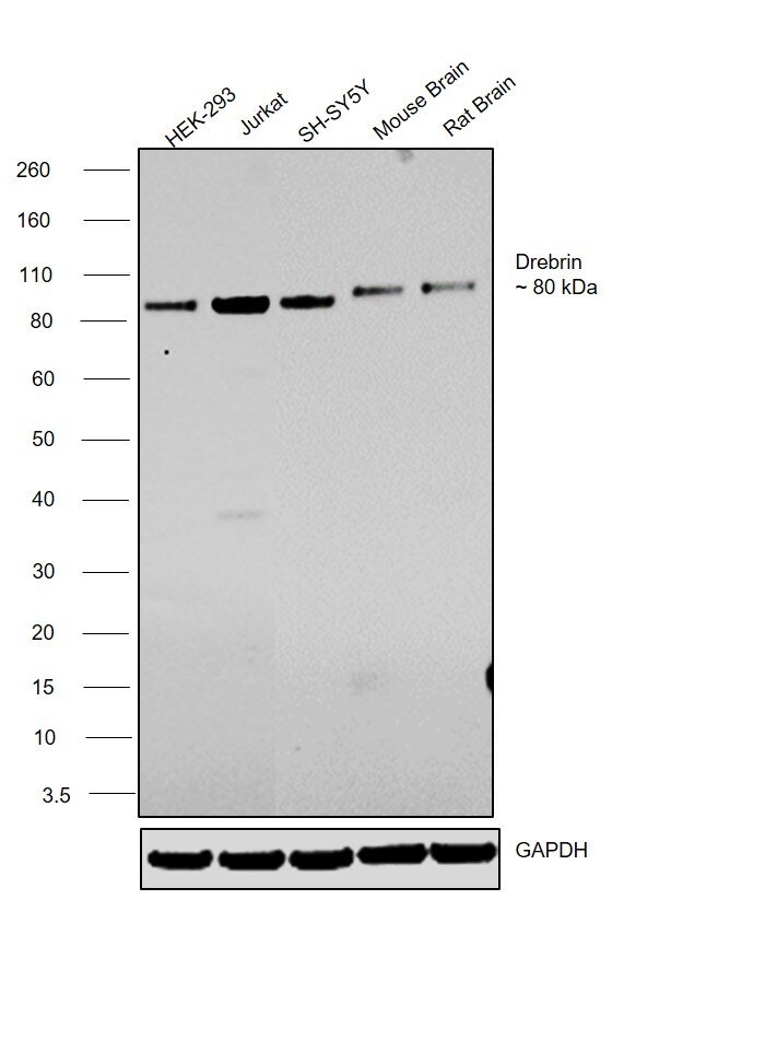
- Western blot was performed using anti-Drebrin Monoclonal Antibody (M2F6) (Product # MA1-20377) and a 80 kDa band corresponding to Drebrin was observed across cell lines and tissues tested. Whole cell extracts (30 µg lysate) of HEK-293 (Lane 1), Jurkat (Lane 2), SH-SY5Y (Lane 3), tissue extracts of Mouse Brain (Lane 4) and Rat Brain (Lane 5) were electrophoresed using Novex® NuPAGE® 12 % Bis-Tris gel (Product # NP0342BOX). Resolved proteins were then transferred onto a nitrocellulose membrane (Product # IB23001) by iBlot® 2 Dry Blotting System (Product # IB21001). The blot was probed with the primary antibody (1:1000 dilution) and detected by chemiluminescence with Goat anti-Mouse IgG (H+L) Superclonal™ Recombinant Secondary Antibody, HRP (Product # A28177, 1:4000 dilution) using the iBright FL 1000 (Product # A32752). Chemiluminescent detection was performed using Novex® ECL Chemiluminescent Substrate Reagent Kit (Product # WP20005).