Explore
 Validate
 Learn
LearnMA1-20832
antibody from Invitrogen Antibodies
Targeting: CENPA
CenH3, CENP-A
 Western blot
 Immunocytochemistry Immunohistochemistry
 Blocking/Neutralizing Chromatin Immunoprecipitation
Antibody data
- Antibody Data
- Antigen structure
- References [1]
- Comments [0]
- Validations
- Immunocytochemistry [3]
- Immunohistochemistry [1]
Submit
Validation data
Reference
Comment
Report error
- Product number
- MA1-20832 - Provider product page

- Provider
- Invitrogen Antibodies
- Product name
- CENPA Monoclonal Antibody (3-19)
- Antibody type
- Monoclonal
- Antigen
- Synthetic peptide
- Description
- Recommended positive controls: , BE cells. This antibody does not cross react with mouse CENPA proteins. Store product as a concentrated solution. Centrifuge briefly prior to opening the vial.
- Reactivity
- Human, Chicken/Avian
- Host
- Mouse
- Isotype
- IgG
- Antibody clone number
- 3-19
- Vial size
- 50 μg
- Concentration
- 1 mg/mL
- Storage
- Store at 4°C short term. For long term storage, store at -20°C, avoiding freeze/thaw cycles.
Submitted references Inheritance of CENP-A Nucleosomes during DNA Replication Requires HJURP.
Zasadzińska E, Huang J, Bailey AO, Guo LY, Lee NS, Srivastava S, Wong KA, French BT, Black BE, Foltz DR
Developmental cell 2018 Nov 5;47(3):348-362.e7
Developmental cell 2018 Nov 5;47(3):348-362.e7
No comments: Submit comment
Supportive validation
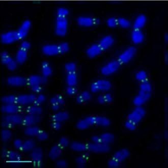
- Submitted by
- Invitrogen Antibodies (provider)
- Main image
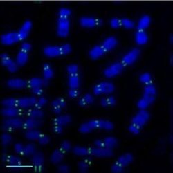
- Experimental details
- Immunofluorescent analysis of CENPA in human metaphase chromosomes using a CENPA monoclonal antibody (Product # MA1-20832) followed by detection using a goat anti-mouse secondary antibody (green).
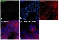
- Submitted by
- Invitrogen Antibodies (provider)
- Main image

- Experimental details
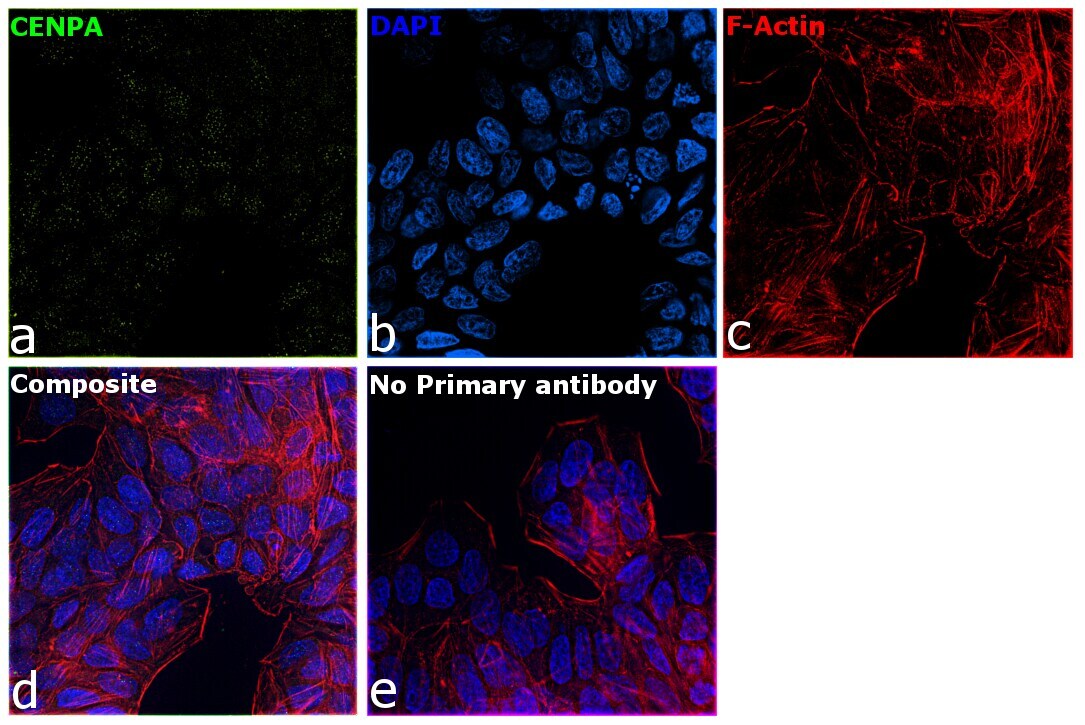
- Immunofluorescence analysis of CENPA was performed using 70% confluent log phase HT-29 cells. The cells were fixed with 4% paraformaldehyde for 10 minutes, permeabilized with 0.1% Triton™ X-100 for 15 minutes, and blocked with 2% BSA for 45 minutes at room temperature. The cells were labeled with CENPA Monoclonal Antibody (3-19) (Product # MA1-20832) at 1:200 in 0.1% BSA, incubated at 4 degree celsius overnight and then labeled with Donkey anti-Mouse IgG (H+L) Highly Cross-Adsorbed Secondary Antibody, Alexa Fluor Plus 488 (Product # A32766), (1:2000), for 45 minutes at room temperature (Panel a: Green). Nuclei (Panel b:Blue) were stained with ProLong™ Diamond Antifade Mountant with DAPI (Product # P36962). F-actin (Panel c: Red) was stained with Rhodamine Phalloidin (Product # R415, 1:300). Panel d represents the merged image showing Centromere localization. Panel e represents control cells with no primary antibody to assess background. The images were captured at 60X magnification.

- Submitted by
- Invitrogen Antibodies (provider)
- Main image
- Experimental details
- Immunofluorescence analysis of CENPA was performed using 70% confluent log phase HT-29 cells. The cells were fixed with 4% paraformaldehyde for 10 minutes, permeabilized with 0.1% Triton™ X-100 for 15 minutes, and blocked with 2% BSA for 45 minutes at room temperature. The cells were labeled with CENPA Monoclonal Antibody (3-19) (Product # MA1-20832) at 1:200 in 0.1% BSA, incubated at 4 degree celsius overnight and then labeled with Donkey anti-Mouse IgG (H+L) Highly Cross-Adsorbed Secondary Antibody, Alexa Fluor Plus 488 (Product # A32766), (1:2000), for 45 minutes at room temperature (Panel a: Green). Nuclei (Panel b:Blue) were stained with ProLong™ Diamond Antifade Mountant with DAPI (Product # P36962). F-actin (Panel c: Red) was stained with Rhodamine Phalloidin (Product # R415, 1:300). Panel d represents the merged image showing Centromere localization. Panel e represents control cells with no primary antibody to assess background. The images were captured at 60X magnification.
Supportive validation
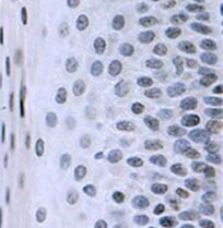
- Submitted by
- Invitrogen Antibodies (provider)
- Main image
- Experimental details
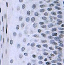
- Immunohistochemical detection of CENP-A on paraffin embedded section of a squamous epithelium of human tonsil using CENPA Monoclonal Antibody (3-19) (Product # MA1-20832).