Explore
 Validate
 Learn
Learn Western blot
 Immunocytochemistry
Antibody data
- Antibody Data
- Antigen structure
- References [1]
- Comments [0]
- Validations
- Western blot [3]
- Other assay [1]
Submit
Validation data
Reference
Comment
Report error
- Product number
- PA1-1042 - Provider product page

- Provider
- Invitrogen Antibodies
- Product name
- Anti-Syntaxin 1 Polyclonal Antibody
- Antibody type
- Polyclonal
- Antigen
- Synthetic peptide
- Description
- PA1-1042 detects Syntaxin 1 in rat, mouse and bovine samples. PA1-1042 has been successfully used in Western blot procedures. By Western blot, this antibody detects a ~33 kDa protein representing Syntaxin 1 in rat, mouse and bovine brain tissue samples. The PA1-1042 immunogen is a synthetic peptide corresponding to residues V(22) T V D R D R F M D E F F E Q V E E I R(41) of rat Syntaxin 1. This sequence is conserved in mouse, human and bovine. The PA1-1042 immunizing peptide (Cat. # PEP-278) is available for use in neutralization and control experiments.
- Reactivity
- Mouse, Rat, Bovine
- Host
- Rabbit
- Isotype
- IgG
- Vial size
- 100 µg
- Concentration
- 1 mg/mL
- Storage
- -20° C, Avoid Freeze/Thaw Cycles
Submitted references Enhanced hippocampal long-term potentiation and fear memory in Btbd9 mutant mice.
DeAndrade MP, Zhang L, Doroodchi A, Yokoi F, Cheetham CC, Chen HX, Roper SN, Sweatt JD, Li Y
PloS one 2012;7(4):e35518
PloS one 2012;7(4):e35518
No comments: Submit comment
Supportive validation
- Submitted by
- Invitrogen Antibodies (provider)
- Main image

- Experimental details
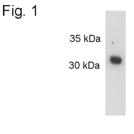
- Western blot detection of Syntaxin 1 from rat brain using Product # PA1-1042.
- Submitted by
- Invitrogen Antibodies (provider)
- Main image
- Experimental details
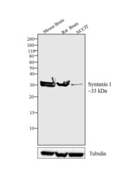
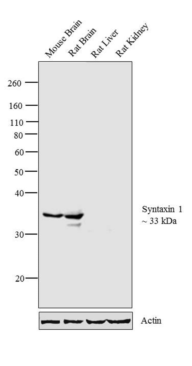
- Western blot analysis was performed on tissue and whole cell extracts (30 µg lysate) of Mouse Brain (Lane 1), Rat Brain (Lane 2) and NCCIT (Lane 3). The blots were probed with Anti-Syntaxin 1 Rabbit Polyclonal Antibody (Product # PA1-1042, 1:5000 dilution) and detected by chemiluminescence using Goat anti-Rabbit IgG (H+L) Superclonal™ Secondary Antibody, HRP conjugate (Product # A27036, 1:2500 dilution). A ~ 33 kDa band corresponding to Syntaxin 1 was observed across cell lines tested. Known quantity of protein samples were electrophoresed using Novex® NuPAGE® 10 % Bis-Tris gel (Product # NP0302BOX), XCell SureLock™ Electrophoresis System (Product # EI0002) and Novex® Sharp Pre-Stained Protein Standard (Product # LC5800). Resolved proteins were then transferred onto a nitrocellulose membrane with iBlot® 2 Dry Blotting System (Product # IB21001). The membrane was probed with the relevant primary and secondary Antibody following blocking with 5 % skimmed milk. Chemiluminescent detection was performed using Pierce™ ECL Western Blotting Substrate (Product # 32106).

- Submitted by
- Invitrogen Antibodies (provider)
- Main image
- Experimental details
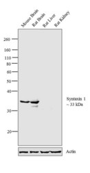
- Western blot analysis was performed on tissue extracts (30 µg lysate) of Mouse Brain (Lane 1), Rat Brain (Lane 2), Rat Liver (Lane 3) and Rat Kidney (Lane 4). The blot was probed with Anti- Syntaxin 1 Rabbit Polyclonal Antibody (Product # PA1-1042, 1:1000 dilution) and detected by chemiluminescence using Goat anti-Rabbit IgG (H+L) Superclonal™ Secondary Antibody, HRP conjugate (Product # A27036, 0.25 µg/mL, 1:4000 dilution). A 33 kDa band corresponding to Syntaxin 1 was observed in Mouse Brain, Rat Brain and not observed in other tissues which are documented to be Syntaxin 1 negative.
Supportive validation
- Submitted by
- Invitrogen Antibodies (provider)
- Main image
- Experimental details
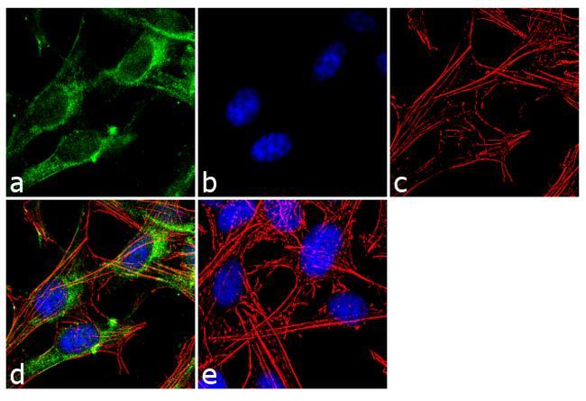
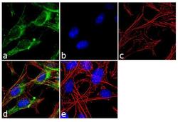
- Immunofluorescence analysis of Syntaxin 1 was performed using 70% confluent log phase SH-SY5Y cells. The cells were fixed with 4% paraformaldehyde for 10 minutes, permeabilized with 0.1% Triton™ X-100 for 10 minutes, and blocked with 2% BSA for 1 hour at room temperature. The cells were labeled with Syntaxin-1 Rabbit Polyclonal Antibody (Product # PA1-1042) at 2 µg/mL in 0.1% BSA and incubated for 3 hours at room temperature and then labeled with Goat anti-Rabbit IgG (H+L) Superclonal™ Secondary Antibody, Alexa Fluor® 488 conjugate (Product # A27034) a dilution of 1:2000 for 45 minutes at room temperature (Panel a: green). Nuclei (Panel b: blue) were stained with SlowFade® Gold Antifade Mountant with DAPI (Product # S36938). F-actin (Panel c: red) was stained with Alexa Fluor® 555 Rhodamine Phalloidin (Product # R415, 1:300). Panel d represents the merged image showing cytoplasmic localization. Panel e shows the no primary antibody control. The images were captured at 60X magnification.