 Explore
 Validate
 Learn
Learn Western blot
 Immunocytochemistry
 Immunohistochemistry
Antibody data
- Antibody Data
- Antigen structure
- References [3]
- Comments [0]
- Validations
- Western blot [1]
- Immunocytochemistry [1]
Submit
Validation data
Reference
Comment
Report error
- Product number
- AMAb90591 - Provider product page

- Provider
- Atlas Antibodies
- Proper citation
- Atlas Antibodies Cat#AMAb90591, RRID:AB_2665598
- Product name
- Anti-RUNX2
- Antibody type
- Monoclonal
- Description
- Monoclonal Antibody against Human RUNX2, Clone ID: CL0232, Gene description: runt-related transcription factor 2, Alternative Gene Names: AML3, CBFA1, CCD, CCD1, PEBP2A1, PEBP2aA1, Validated applications: WB, IHC, ICC, Uniprot ID: Q13950, Storage: Store at +4°C for short term storage. Long time storage is recommended at -20°C.
- Reactivity
- Human
- Host
- Mouse
- Conjugate
- Unconjugated
- Isotype
- IgG
- Antibody clone number
- CL0232
- Vial size
- 100 µl
- Concentration
- 1.0 mg/ml
- Storage
- Store at +4°C for short term storage. Long time storage is recommended at -20°C.
- Handling
- The antibody solution should be gently mixed before use.
Submitted references Synergistic Effects of Photobiomodulation and Differentiation Inducers on Osteogenic Differentiation of Adipose-Derived Stem Cells in Three-Dimensional Culture
Proteoglycan 4 Modulates Osteogenic Smooth Muscle Cell Differentiation during Vascular Remodeling and Intimal Calcification
Beneficial Effects of Galectin-3 Blockade in Vascular and Aortic Valve Alterations in an Experimental Pressure Overload Model
Da Silva D, Crous A, Abrahamse H
International Journal of Molecular Sciences 2024;25(24):13350
International Journal of Molecular Sciences 2024;25(24):13350
Proteoglycan 4 Modulates Osteogenic Smooth Muscle Cell Differentiation during Vascular Remodeling and Intimal Calcification
Seime T, Akbulut A, Liljeqvist M, Siika A, Jin H, Winski G, van Gorp R, Karlöf E, Lengquist M, Buckler A, Kronqvist M, Waring O, Lindeman J, Biessen E, Maegdefessel L, Razuvaev A, Schurgers L, Hedin U, Matic L
Cells 2021;10(6):1276
Cells 2021;10(6):1276
Beneficial Effects of Galectin-3 Blockade in Vascular and Aortic Valve Alterations in an Experimental Pressure Overload Model
Ibarrola J, Martínez-Martínez E, Sádaba J, Arrieta V, García-Peña A, Álvarez V, Fernández-Celis A, Gainza A, Rossignol P, Cachofeiro Ramos V, López-Andrés N
International Journal of Molecular Sciences 2017;18(8):1664
International Journal of Molecular Sciences 2017;18(8):1664
No comments: Submit comment
Enhanced validation
- Submitted by
- Atlas Antibodies (provider)
- Enhanced method
- Genetic validation
- Main image
- Experimental details
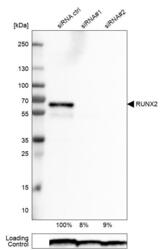
- Western blot analysis in U-251MG cells transfected with control siRNA, target specific siRNA probe #1 and #2, using Anti-RUNX2 antibody. Remaining relative intensity is presented. Loading control: Anti-PPIB.
- Sample type
- Human
- Protocol
- Protocol

Supportive validation
- Submitted by
- Atlas Antibodies (provider)
- Main image
- Experimental details
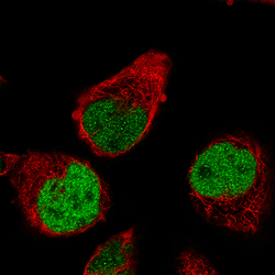
- Immunofluorescence staining of RH-30 cells using the Anti-RUNX2 monoclonal antibody, showing specific staining in the nucleoplasm in green. Microtubule- and nuclear probes are visualized in red and blue, respectively (where available).
- Sample type
- Human
