Explore
 Validate
 Learn
Learn Western blot
 ELISA
ELISAAntibody data
- Antibody Data
- Antigen structure
- References [3]
- Comments [0]
- Validations
- Western blot [1]
Submit
Validation data
Reference
Comment
Report error
- Product number
- 32-1000 - Provider product page

- Provider
- Invitrogen Antibodies
- Product name
- EAAC1 Monoclonal Antibody (35-A9)
- Antibody type
- Monoclonal
- Antigen
- Synthetic peptide
- Description
- Immunogen sequence: KETDSARSR SAPMSPSDFL DKLMGRTSGY DARIRPNFKG PPVNVTCNIF INSFGSIAET TMDYRVNIFL RQKWNDPRLA YSEYPDDSLD LDPSMLDSIW KPDLFFANEK GANFHEVTTD NKLLRIFKNG NVLYSIRLTL TLSCPMDLKN FPMDVQTCIM QLESFGYTMN DLIFEWQDEA PVQVAEGLTL PQFLLKEEKD LRYCTKHYNT GKFTCIEVRF HLERQMGY (29-255 aa encoded by BC036086 )
- Reactivity
- Human, Rat
- Host
- Mouse
- Isotype
- IgG
- Antibody clone number
- 35-A9
- Vial size
- 100 µg
- Concentration
- 0.5 mg/mL
- Storage
- -20°C
Submitted references In vitro Ag Cross-presentation and in vivo Ag Cross-presentation by Dendritic Cells in the Mouse.
Impaired spinal cord glutamate transport capacity and reduced sensitivity to riluzole in a transgenic superoxide dismutase mutant rat model of amyotrophic lateral sclerosis.
Immunohistochemical localization of the neuron-specific glutamate transporter EAAC1 (EAAT3) in rat brain and spinal cord revealed by a novel monoclonal antibody.
Ghosh M, Shapiro LH
Bio-protocol 2012 Dec 20;2(24):e305
Bio-protocol 2012 Dec 20;2(24):e305
Impaired spinal cord glutamate transport capacity and reduced sensitivity to riluzole in a transgenic superoxide dismutase mutant rat model of amyotrophic lateral sclerosis.
Dunlop J, Beal McIlvain H, She Y, Howland DS
The Journal of neuroscience : the official journal of the Society for Neuroscience 2003 Mar 1;23(5):1688-96
The Journal of neuroscience : the official journal of the Society for Neuroscience 2003 Mar 1;23(5):1688-96
Immunohistochemical localization of the neuron-specific glutamate transporter EAAC1 (EAAT3) in rat brain and spinal cord revealed by a novel monoclonal antibody.
Shashidharan P, Huntley GW, Murray JM, Buku A, Moran T, Walsh MJ, Morrison JH, Plaitakis A
Brain research 1997 Oct 31;773(1-2):139-48
Brain research 1997 Oct 31;773(1-2):139-48
No comments: Submit comment
Supportive validation
- Submitted by
- Invitrogen Antibodies (provider)
- Main image

- Experimental details
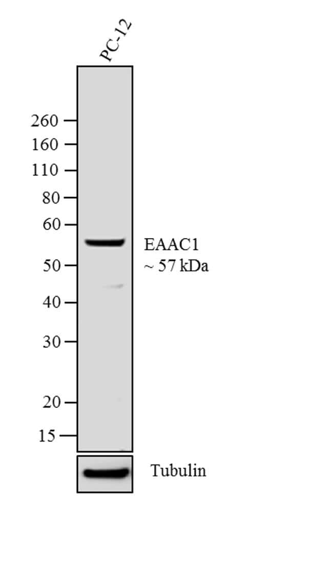
- Western blot analysis was performed on whole cell extracts of PC-12 (Lane 1). The blot was probed with Anti-EAAC1 (Glutamate Transporter) Mouse Monoclonal Antibody (Product # 32-1000, 2 µg/mL) and detected by chemiluminescence using Goat anti-Mouse IgG (H+L) Superclonal™ Secondary Antibody, HRP conjugate (Product # A28177, 0.4 µg/mL, 1:2500 dilution). A 57 kDa band corresponding to EAAC1 was observed in the cell line tested. Known quantity of protein samples were electrophoresed using Novex® NuPAGE®12% Bis-Tris gel (Product # NP0342BOX), XCell SureLock™ Electrophoresis System (Product # EI0002) and Novex® Sharp Pre-Stained Protein Standard (Product # LC5800). Resolved proteins were then transferred onto a nitrocellulose membrane with iBlot® 2 Dry Blotting System (Product # IB21001). The membrane was probed with the relevant primary and secondary Antibody. Chemiluminescent detection was performed using Pierce™ ECL Western Blotting Substrate (Product # 32106).