Explore
 Validate
 Learn
Learn Western blot
 Immunoprecipitation
Antibody data
- Antibody Data
- Antigen structure
- References [1]
- Comments [0]
- Validations
- Western blot [1]
- Immunocytochemistry [1]
- Flow cytometry [1]
Submit
Validation data
Reference
Comment
Report error
- Product number
- MA5-16944 - Provider product page

- Provider
- Invitrogen Antibodies
- Product name
- CD10 Monoclonal Antibody (SN5c)
- Antibody type
- Monoclonal
- Antigen
- Purifed from natural sources
- Description
- For FACS analysis, use 10 µL of the suggested working dilution to label 1x10^6 cells in 100 µL.
- Reactivity
- Human
- Host
- Mouse
- Isotype
- IgG
- Antibody clone number
- SN5c
- Vial size
- 100 µg
- Concentration
- 1 mg/mL
- Storage
- Store at 4°C short term. For long term storage, store at -20°C, avoiding freeze/thaw cycles.
Submitted references LECT2 drives haematopoietic stem cell expansion and mobilization via regulating the macrophages and osteolineage cells.
Lu XJ, Chen Q, Rong YJ, Yang GJ, Li CH, Xu NY, Yu CH, Wang HY, Zhang S, Shi YH, Chen J
Nature communications 2016 Sep 6;7:12719
Nature communications 2016 Sep 6;7:12719
No comments: Submit comment
Supportive validation
- Submitted by
- Invitrogen Antibodies (provider)
- Main image
- Experimental details
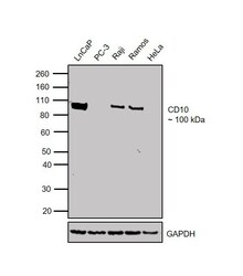
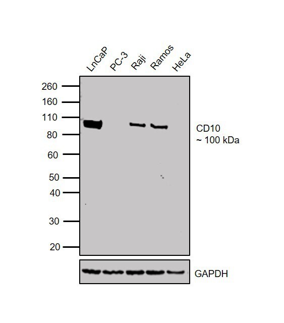
- Western blot was performed using Anti-CD10 Mouse Monoclonal Antibody (Product # MA5-16944) and a 100 kDa band corresponding to CD10 was observed in cell lines tested except for PC-3 and HeLa which are reported to be negative. Membrane enriched extracts (30 µg lysate) of LNCaP (Lane 1), PC-3 (Lane 2), Raji (Lane 3), Ramos (Lane 4) and HeLa (Lane 5) were electrophoresed using Novex® NuPAGE® 4-12% Bis-Tris Protein Gel (Product # NP0322BOX). Resolved proteins were then transferred onto a nitrocellulose membrane (Product # IB23001) by iBlot® 2 Dry Blotting System (Product # IB21001). The blot was probed with the primary antibody (1:1000 dilution) and detected by chemiluminescence with Goat anti-Mouse IgG (H+L) Superclonal™ Recombinant Secondary Antibody, HRP (Product # A28177, 1:4000 dilution) using the iBright FL 1000 (Product # A32752). Chemiluminescent detection was performed using Novex® ECL Chemiluminescent Substrate Reagent Kit (Product # WP20005).
Supportive validation
- Submitted by
- Invitrogen Antibodies (provider)
- Main image
- Experimental details
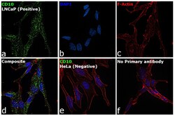
- Immunofluorescence analysis of CD10 was performed using LNCaP and HeLa cells. The cells were fixed with 4% paraformaldehyde for 10 minutes, permeabilized with 0.1% Triton™ X-100 for 15 minutes, and blocked with 2% BSA for 1 hour at room temperature. The cells were labeled with CD10 Mouse Monoclonal Antibody (Product # MA5-16944) at 1:100 dilution in 0.1% BSA and incubated overnight at 4 degree and then labeled with Goat anti-Mouse IgG (H+L) Highly Cross-Adsorbed Secondary Antibody, Alexa Fluor Plus 488 (Product # A32723) at a dilution of 1:2000 for 45 minutes at room temperature (Panel a: green) in LNCaP cells. Nuclei (Panel b: blue) were stained with ProLong™ Diamond Antifade Mountant with DAPI (Product # P36962). F-actin (Panel c: red) was stained with Rhodamine Phalloidin (Product # R415, 1:300). Panel d represents the merged image of LNCaP cells, which is a positive model for CD10 expression showing localization of CD10 cytoplasmic domain around plasma membrane. Panel e represents the merged image of HeLa cells, that are null for CD10 protein expression. Panel f represents control cells with no primary antibody to assess background. The images were captured at 60X magnification.

Supportive validation
- Submitted by
- Invitrogen Antibodies (provider)
- Main image

- Experimental details
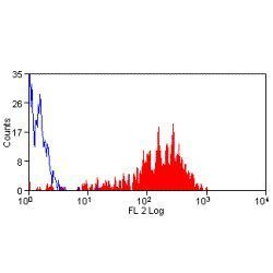
- Flow cytometric analysis of thrombin activated human peripheral blood platelets with Mouse anti Human CD62P conjugated to RPE