Explore
 Validate
 Learn
Learn Western blot
Antibody data
- Antibody Data
- Antigen structure
- References [0]
- Comments [0]
- Validations
- Western blot [6]
- Immunohistochemistry [2]
Submit
Validation data
Reference
Comment
Report error
- Product number
- PA5-29354 - Provider product page

- Provider
- Invitrogen Antibodies
- Product name
- CD10 Polyclonal Antibody
- Antibody type
- Polyclonal
- Antigen
- Recombinant protein fragment
- Description
- Recommended positive controls: Jurkat, mouse kidney, rat kidney. Predicted reactivity: Mouse (91%), Rat (93%), Dog (89%), Rabbit (88%), Rhesus Monkey (98%), Bovine (86%). Store product as a concentrated solution. Centrifuge briefly prior to opening the vial.
- Reactivity
- Human, Mouse, Rat
- Host
- Rabbit
- Isotype
- IgG
- Vial size
- 100 µL
- Concentration
- 1.01 mg/mL
- Storage
- Store at 4°C short term. For long term storage, store at -20°C, avoiding freeze/thaw cycles.
No comments: Submit comment
Supportive validation
- Submitted by
- Invitrogen Antibodies (provider)
- Main image

- Experimental details
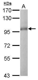
- Western blot analysis of CD10/MME using 30 µg of Jurkat lysate. Samples were loaded onto a 7.5% SDS-PAGE gel and probed with a CD10/MME polyclonal antibody (Product # PA5-29354) at a dilution of 1:1000.

- Submitted by
- Invitrogen Antibodies (provider)
- Main image

- Experimental details
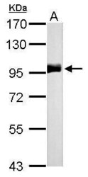
- Western blot analysis of CD10/MME using 50 µg mouse kidney lysate. Samples were loaded onto a 7.5% SDS-PAGE gel and probed with a CD10/MME polyclonal antibody (Product # PA5-29354) at a dilution of 1:500.
- Submitted by
- Invitrogen Antibodies (provider)
- Main image
- Experimental details
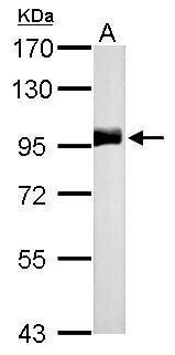
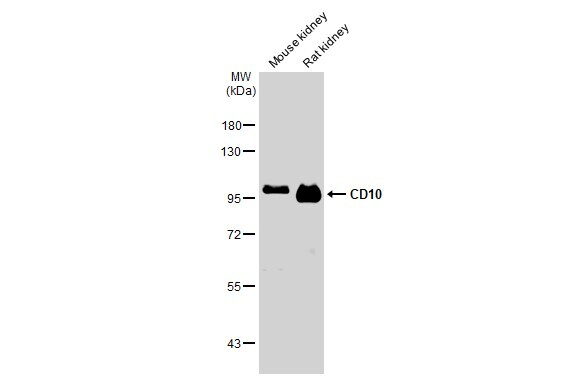
- CD10 Polyclonal Antibody detects MME protein by western blot analysis. A. 50 µg rat kidney lysate/extract.7.5% SDS-PAGE. CD10 Polyclonal Antibody (Product # PA5-29354) dilution: 1:500. The HRP-conjugated anti-rabbit IgG antibody was used to detect the primary antibody.
- Submitted by
- Invitrogen Antibodies (provider)
- Main image

- Experimental details
- Western Blot using CD10 Polyclonal Antibody (Product # PA5-29354). Various tissue extracts (50 µg) were separated by 7.5% SDS-PAGE, and the membrane was blotted with CD10 Polyclonal Antibody (Product # PA5-29354) diluted at 1:500. The HRP-conjugated anti-rabbit IgG antibody was used to detect the primary antibody.
- Submitted by
- Invitrogen Antibodies (provider)
- Main image

- Experimental details
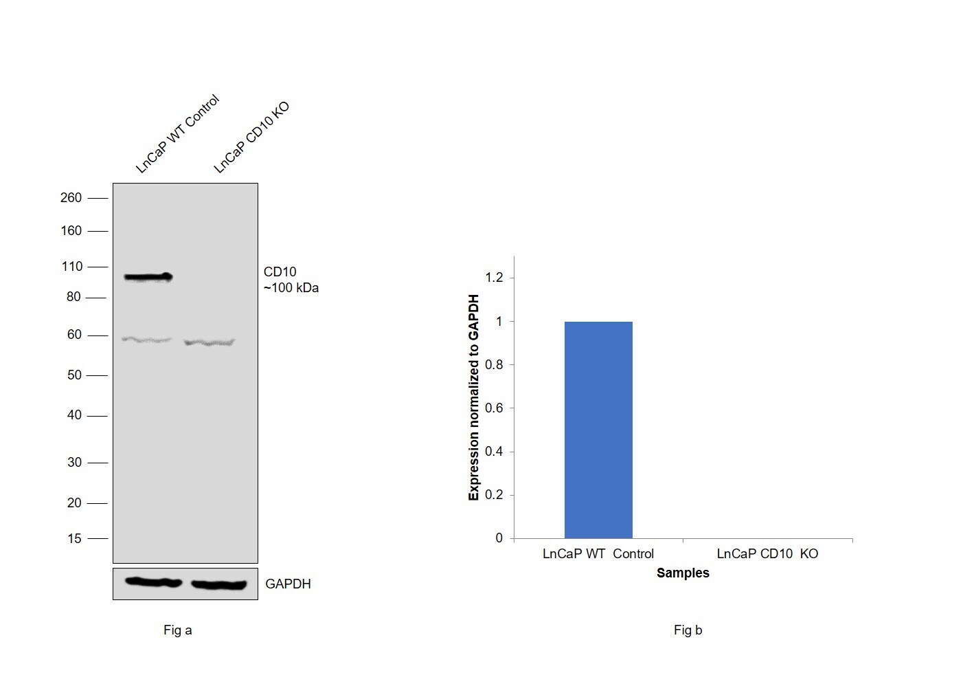
- Knockout of CD10 was achieved by CRISPR-Cas9 genome editing using LentiArray™ Lentiviral sgRNA (Product # A32042, AssayID CRISPR1056041_LV) and LentiArray Cas9 Lentivirus (Product # A32064). Western blot analysis of CD10 was performed by loading 30 µg of LNCaP wild type (Lane 1) and LNCaP CD10 KO (Lane 2) membrane extracts. The samples were electrophoresed using Novex® NuPAGE® 4-12% Bis-Tris Protein Gel (Product # NP0321BOX). Resolved proteins were then transferred onto a nitrocellulose membrane (Product # IB23001) by iBlot® 2 Dry Blotting System (Product # IB21001). The blot was probed with Anti-CD10 Polyclonal Antibody (Product # PA5-29354) using 0.5 µg/mL dilution and Goat anti-Rabbit IgG (H+L), Superclonal™ Recombinant Secondary Antibody, HRP (Product # A27036, 1:4000 dilution) using the iBright FL 1000 (Product # A32752). Chemiluminescent detection was performed using Novex® ECL Chemiluminescent Substrate Reagent Kit (Product # WP20005). Loss of signal upon CRISPR mediated knockout (KO) using the LentiArray™ CRISPR product line confirms that antibody is specific to CD10. An uncharacterized band was observed at ~60 kDa in both the samples.
- Submitted by
- Invitrogen Antibodies (provider)
- Main image
- Experimental details
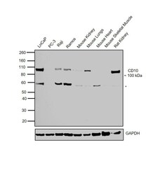
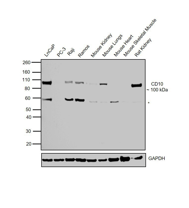
- Western blot was performed using Anti-CD10 Rabbit Polyclonal Antibody (Product # PA5-29354) and a 100 kDa band corresponding to CD10 was observed in cell lines and tissues tested except for PC-3, Mouse Heart and Mouse Skeletal Muscle. Membrane enriched extracts (30 µg lysate) of LNCaP (Lane 1), PC-3 (Lane 2), Raji (Lane 3), Ramos (Lane 4), Mouse Kidney (Lane 5), Mouse Lungs (Lane 6), Mouse Heart (Lane 7), Mouse Skeletal Muscle (Lane 8) and Rat Kidney (Lane 9) were electrophoresed using Novex® NuPAGE® 4-12% Bis-Tris Protein Gel (Product # NP0322BOX). Resolved proteins were then transferred onto a nitrocellulose membrane (Product # IB23001) by iBlot® 2 Dry Blotting System (Product # IB21001). The blot was probed with the primary antibody (0.5 µg/mL) and detected by chemiluminescence with Goat anti-Rabbit IgG (H+L) Superclonal™ Recombinant Secondary Antibody, HRP (Product # A27036, 1:4000 dilution) using the iBright FL 1000 (Product # A32752). Chemiluminescent detection was performed using Novex® ECL Chemiluminescent Substrate Reagent Kit (Product # WP20005). An uncharacterized band (*) of 60 kDa was observed across cell lines and tissues.
Supportive validation
- Submitted by
- Invitrogen Antibodies (provider)
- Main image
- Experimental details
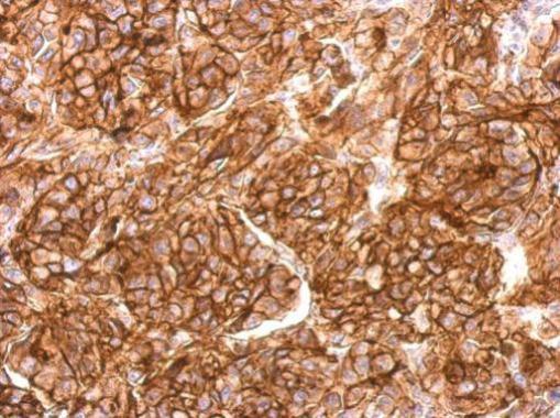
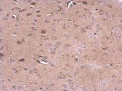
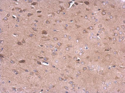
- CD10 Polyclonal Antibody detects CD10 protein at membrane on mouse fore brain by immunohistochemical analysis. Sample: Paraffin-embedded mouse fore brain. CD10 Polyclonal Antibody (Product # PA5-29354) dilution: 1:500. Antigen Retrieval: EDTA based buffer, pH 8.0, 15 min.
- Submitted by
- Invitrogen Antibodies (provider)
- Main image
- Experimental details
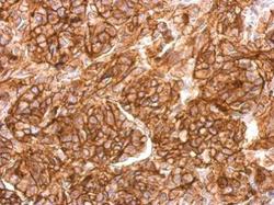
- CD10 Polyclonal Antibody detects MME protein at cytosol and membrane on SkHep1 xenograft by immunohistochemical analysis. Sample: Paraffin-embedded SkHep1 xenograft. CD10 Polyclonal Antibody (Product # PA5-29354) dilution: 1:500. Antigen Retrieval: EDTA based buffer, pH 8.0, 15 min.