Explore
 Validate
 Learn
Learn Western blot
 Immunohistochemistry
Antibody data
- Antibody Data
- Antigen structure
- References [0]
- Comments [0]
- Validations
- Immunohistochemistry [5]
- Flow cytometry [1]
Submit
Validation data
Reference
Comment
Report error
- Product number
- NBP2-15197-0.1mg - Provider product page

- Provider
- Novus Biologicals
- Product name
- Mouse Monoclonal Melan-A/MART-1 Antibody
- Antibody type
- Monoclonal
- Description
- Protein G purified. This antibody recognizes a protein doublet of 20-22kDa, identified as MART-1 (Melanoma Antigen Recognized by T cells 1) or Melan-A. MART-1 is a newly identified melanocyte differentiation antigen recognized by autologous cytotoxic T lymphocytes. Seven other melanoma associated antigens recognized by autologous cytotoxic T cells include MAGE-1, MAGE-3, tyrosinase, gp100, gp75, BAGE-1, and GAGE-1. Subcellular fractionation shows that MART-1 is present in melanosomes and endoplasmic reticulum. This MAb labels melanomas and other tumors showing melanocytic differentiation. It is also a useful positive-marker for angiomyolipomas. It does not stain tumor cells of epithelial, lymphoid, glial, or mesenchymal origin.
- Reactivity
- Human, Mouse, Rat
- Host
- Mouse
- Isotype
- IgG
- Vial size
- 0.1 mg
- Concentration
- 0.2 mg/ml
- Storage
- Store at 4C.
No comments: Submit comment
Supportive validation
- Submitted by
- Novus Biologicals (provider)
- Main image

- Experimental details
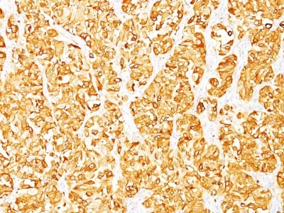
- Immunohistochemistry-Paraffin: Melan-A/MART-1 Antibody (M2-7C10) [NBP2-15197] - Formalin-fixed, paraffin-embedded human skin stained with MART-1 Monoclonal Antibody (M2-7C10).

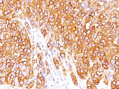
- Submitted by
- Novus Biologicals (provider)
- Main image
- Experimental details
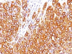
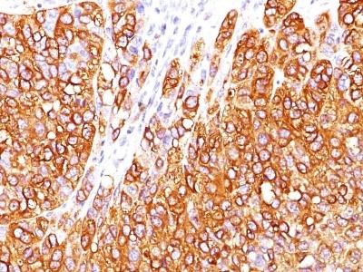
- Immunohistochemistry-Paraffin: Melan-A/MART-1 Antibody (M2-7C10) [NBP2-15197] - Formalin-paraffin human melanoma stained with MART-1 Ab (M2-7C10). Note cytoplasmic staining of cells.
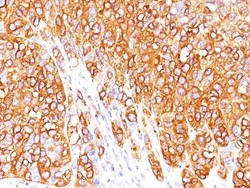
- Submitted by
- Novus Biologicals (provider)
- Main image
- Experimental details
- Immunohistochemistry-Paraffin: Melan-A/MART-1 Antibody (M2-7C10) [NBP2-15197] - Formalin-paraffin human melanoma stained with MART-1 Ab (M2-7C10). Note cytoplasmic staining of cells.
- Submitted by
- Novus Biologicals (provider)
- Main image
- Experimental details
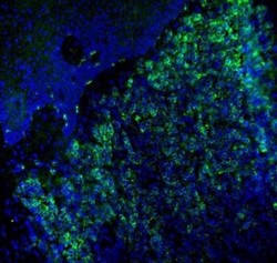
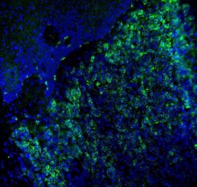
- Immunohistochemistry-Frozen: Melan-A/MART-1 Antibody (M2-7C10) [NBP2-15197] - Analysis using the Alexa-Fluor(R)-488 conjugate for this antibody. Melan-A (green) was detected in human skin (melanoma) using Melan-A-AlexaFluor488 antibody (1:40; 1 hour) in PBS. Nuclei were stained with DAPI (blue). Tissue was fixed in acetone. Image from a verified customer review.
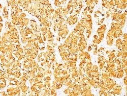
- Submitted by
- Novus Biologicals (provider)
- Main image
- Experimental details
- Immunohistochemistry-Paraffin: Melan-A/MART-1 Antibody (M2-7C10) [NBP2-15197] - Formalin-fixed, paraffin-embedded human Melanoma stained with Melan-A/MART-1 Antibody (M2-7C10).
Supportive validation
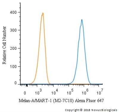
- Submitted by
- Novus Biologicals (provider)
- Main image

- Experimental details
- Flow Cytometry: Melan-A/MART-1 Antibody (M2-7C10) [NBP2-15197] - Flow Cytometry: Melan-A/MART-1 Antibody (M2-7C10) [Alexa Fluor® 647] [NBP2-33148AF647] - An intracellular stain was performed on SK-MEL-28 cells with Melan-A/MART-1 Antibody (M2-7C10) NBP2-33148AF647 (blue) and a matched isotype control (orange). Cells were fixed with 4% PFA and then permeablized with 0.1% saponin. Cells were incubated in an antibody dilution of 2.5 ug/mL for 30 minutes at room temperature. Both antibodies were conjugated to Alexa Fluor 647. Image using the Alexa Fluor 647 form of this antibody.