Explore
 Validate
 Learn
Learn Western blot
 ELISA
ELISAAntibody data
- Antibody Data
- Antigen structure
- References [1]
- Comments [0]
- Validations
- Western blot [1]
- Immunohistochemistry [2]
- Flow cytometry [2]
Submit
Validation data
Reference
Comment
Report error
- Product number
- NBP2-46603 - Provider product page

- Provider
- Novus Biologicals
- Product name
- Mouse Monoclonal Melan-A/MART-1 Antibody
- Antibody type
- Monoclonal
- Description
- Protein G purified.
- Reactivity
- Human, Mouse, Rat
- Host
- Mouse
- Isotype
- IgG
- Vial size
- 0.1 ml
- Concentration
- 1.0 mg/ml
- Storage
- Store at 4C short term. Aliquot and store at -20C long term. Avoid freeze-thaw cycles.
Submitted references Expression of natural killer cell regulatory microRNA by uveal melanoma cancer stem cells.
Joshi P, Kooshki M, Aldrich W, Varghai D, Zborowski M, Singh AD, Triozzi PL
Clinical & experimental metastasis 2016 Dec;33(8):829-838
Clinical & experimental metastasis 2016 Dec;33(8):829-838
No comments: Submit comment
Supportive validation
- Submitted by
- Novus Biologicals (provider)
- Main image
- Experimental details
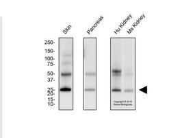
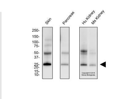
- Western Blot: Melan-A/MART-1 Antibody (A103) [NBP2-46603] - Total protein from human skin, pancreas and kidney and mouse kidney was separated on a 4-20% gel by SDS-PAGE, transferred to PVDF membrane and blocked in 5% non-fat milk in TBST. The membrane was probed with 2.0 ug/ml anti-Melan-A in 1% non-fat milk in TBST and detected with an anti-mouse HRP secondary antibody using chemiluminescence.
Supportive validation
- Submitted by
- Novus Biologicals (provider)
- Main image
- Experimental details
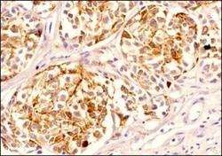
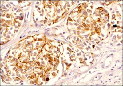
- Immunohistochemistry-Paraffin: Melan-A/MART-1 Antibody (A103) [NBP2-46603] - Analysis of FFPE tissue section of Human Skin Cancer/Melanoma using Melan-A/MART-1 antibody (A103) at 1:100 dilution. The antibody depicted very specific staining which was cytoplasmic with strong punctate appearance in peri-nuclear regions (ER and Golgi) of most of the cancerous cells. The tumor stroma cells did not display any Melan-A/MART-1 expression signal.
- Submitted by
- Novus Biologicals (provider)
- Main image

- Experimental details
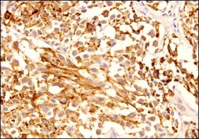
- Immunohistochemistry-Paraffin: Melan-A/MART-1 Antibody (A103) [NBP2-46603] - Analysis of FFPE tissue section of Human Skin Cancer/Melanoma using Melan-A/MART-1 antibody (A103) at 1:100 dilution. The antibody depicted very specific staining which was cytoplasmic with strong punctate appearance in peri-nuclear regions (ER and Golgi) of most of the cancerous cells and membranous-cytoplasmic pattern in some necrotic areas.
Supportive validation
- Submitted by
- Novus Biologicals (provider)
- Main image
- Experimental details
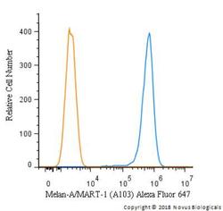
- Flow Cytometry: Melan-A/MART-1 Antibody (A103) [NBP2-46603] - An intracellular stain was performed on SK-MEL-28 cells with Melan-A/MART-1 [A103] Antibody NBP2-46603AF647 (blue) and a matched isotype control (orange). Cells were fixed with 4% PFA and then permeabilized with 0.1% saponin. Cells were incubated in an antibody dilution of 2.5 ug/mL for 30 minutes at room temperature. Both antibodies were conjugated to Alexa Fluor 647.
- Submitted by
- Novus Biologicals (provider)
- Main image
- Experimental details
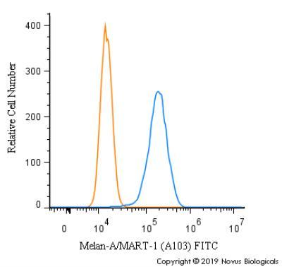
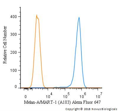
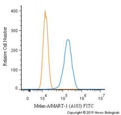
- Flow Cytometry: Melan-A/MART-1 Antibody (A103) [NBP2-46603] - An intracellular stain was performed on SK-MEL-28 cells with Melan-A/MART-1 Antibody [A103] NBP2-46603F (blue) and a matched isotype control (orange). Cells were fixed with 4% PFA and then permeabilized with 0.1% saponin. Cells were incubated in an antibody dilution of 5 ug/mL for 30 minutes at room temperature. Both antibodies were conjugated to FITC.