Explore
 Validate
 Learn
Learn Western blot
 ELISA
ELISAAntibody data
- Antibody Data
- Antigen structure
- References [4]
- Comments [0]
- Validations
- Western blot [1]
- Flow cytometry [1]
Submit
Validation data
Reference
Comment
Report error
- Product number
- ABIN953322 - Provider product page

- Provider
- antibodies-online
- Product name
- anti-Melan A (MLANA) (AA 41-68), (Middle Region) antibody
- Antibody type
- Polyclonal
- Antigen
- KLH conjugated synthetic peptide between 41-68 amino acids from the Central region of human MART-1 Genename: MLANA
- Description
- Protein A column, followed by peptide affinity purification
- Reactivity
- Human
- Host
- Rabbit
- Epitope
- AA 41-68,Middle Region
- Vial size
- 0.4 mL
- Concentration
- 0.25 mg/mL
- Storage
- Store undiluted at 2-8°C for one month or (in aliquots) at -20°C for longer.
- Handling
- Avoid repeated freezing and thawing.
Submitted references Structural basis for the presentation of tumor-associated MHC class II-restricted phosphopeptides to CD4+ T cells.
Melan-a-positive "pseudomelanocytic nests": a pitfall in the histopathologic and immunohistochemical diagnosis of pigmented lesions on sun-damaged skin.
Pigmentation-related genes and their implication in malignant melanoma susceptibility.
The ocular albinism type 1 (OA1) G-protein-coupled receptor functions with MART-1 at early stages of melanogenesis to control melanosome identity and composition.
Li Y, Depontieu FR, Sidney J, Salay TM, Engelhard VH, Hunt DF, Sette A, Topalian SL, Mariuzza RA
Journal of molecular biology 2010 Jun 18;399(4):596-603
Journal of molecular biology 2010 Jun 18;399(4):596-603
Melan-a-positive "pseudomelanocytic nests": a pitfall in the histopathologic and immunohistochemical diagnosis of pigmented lesions on sun-damaged skin.
Beltraminelli H, Shabrawi-Caelen LE, Kerl H, Cerroni L
The American Journal of dermatopathology 2009 May;31(3):305-8
The American Journal of dermatopathology 2009 May;31(3):305-8
Pigmentation-related genes and their implication in malignant melanoma susceptibility.
Fernandez LP, Milne RL, Pita G, Floristan U, Sendagorta E, Feito M, Avilés JA, Martin-Gonzalez M, Lázaro P, Benítez J, Ribas G
Experimental dermatology 2009 Jul;18(7):634-42
Experimental dermatology 2009 Jul;18(7):634-42
The ocular albinism type 1 (OA1) G-protein-coupled receptor functions with MART-1 at early stages of melanogenesis to control melanosome identity and composition.
Giordano F, Bonetti C, Surace EM, Marigo V, Raposo G
Human molecular genetics 2009 Dec 1;18(23):4530-45
Human molecular genetics 2009 Dec 1;18(23):4530-45
No comments: Submit comment
Supportive validation
- Submitted by
- antibodies-online (provider)
- Main image
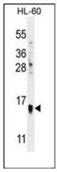
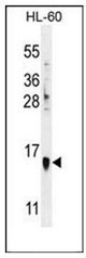
- Experimental details
- Western blot analysis of MART-1 Antibody (Center) Cat.-No AP52610PU-N in HL-60 cell line lysates (35ug/lane). This demonstrates the MART-1 antibody detected the MART-1/Melan-A protein (arrow).
Supportive validation
- Submitted by
- antibodies-online (provider)
- Main image
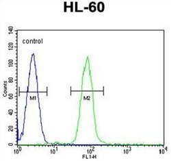
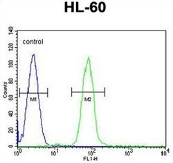
- Experimental details
- Flow cytometric analysis of HL-60 cells using MART-1 Antibody (Center) Cat.-No AP52610PU-N (right histogram) compared to a negative control cell (left histogram). FITC-conjugated goat-anti-rabbit secondary antibodies were used for the analysis.