 Explore
 Validate
 Learn
Learn Western blot
Antibody data
- Antibody Data
- Antigen structure
- References [0]
- Comments [0]
- Validations
- Western blot [1]
- Immunohistochemistry [2]
- Other assay [1]
Submit
Validation data
Reference
Comment
Report error
- Product number
- V2290 - Provider product page

- Provider
- NSJ Bioreagents
- Product name
- Tyrosinase Antibody
- Antibody type
- Monoclonal
- Description
- This highly specific Tyrosinase antibody is suitable for use in Immunohistochemistry applications with human samples.
- Reactivity
- Human
- Host
- Mouse
- Conjugate
- Unconjugated
- Antibody clone number
- T311
- Vial size
- 20 ug (with BSA and sodium azide), 100 ug (with BSA and sodium azide), 100 ug (without BSA or sodium azide), 7 ml IHC only format (if applicable)
- Concentration
- 0.2 mg/ml, 1 mg/ml
- Storage
- Store the Tyrosinase antibody at 2-8oC (with azide) or aliquot and store at -20oC or colder (without azide).
No comments: Submit comment
Supportive validation
- Submitted by
- NSJ Bioreagents (provider)
- Main image

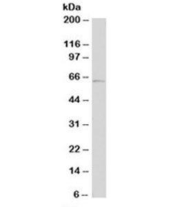
- Experimental details
- Western blot testing of human A431 cell lysate with Tyrosinase antibody (clone T311). Expected molecular weight: ~60-84kDa depending on glycosylation level.
Supportive validation
- Submitted by
- NSJ Bioreagents (provider)
- Main image

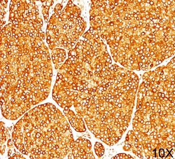
- Experimental details
- IHC staining of melanoma tissue (10X) with Tyrosinase antibody (T311).
- Submitted by
- NSJ Bioreagents (provider)
- Main image
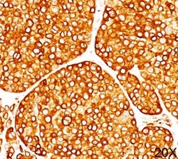
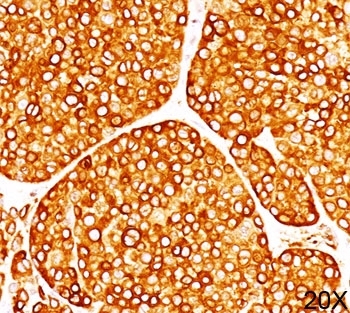
- Experimental details
- IHC staining of melanoma tissue (20X) with Tyrosinase antibody (T311).
Supportive validation
- Submitted by
- NSJ Bioreagents (provider)
- Main image
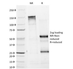
- Experimental details
- SDS-PAGE Analysis of Purified, BSA-Free Tyrosinase Antibody (clone T311). Confirmation of Integrity and Purity of the Antibody.
