Explore
 Validate
 Learn
Learn Western blot
 Immunocytochemistry
Antibody data
- Antibody Data
- Antigen structure
- References [1]
- Comments [0]
- Validations
- Immunocytochemistry [2]
- Immunoprecipitation [1]
- Immunohistochemistry [1]
- Other assay [1]
Submit
Validation data
Reference
Comment
Report error
- Product number
- PA5-34682 - Provider product page

- Provider
- Invitrogen Antibodies
- Product name
- Cyclin A2 Polyclonal Antibody
- Antibody type
- Polyclonal
- Antigen
- Recombinant full-length protein
- Description
- Recommended positive controls: 293T, A431, HeLa, HepG2, IMR32, SK-N-AS, HeLa(100 µg/mL Bleomycin treatment for 24 hr), Neuro2A, PC-12, PC3. Predicted reactivity: Rat (80%), Pig (89%), Rabbit (84%), Rhesus Monkey (98%), Bovine (84%). Store product as a concentrated solution. Centrifuge briefly prior to opening the vial.
- Reactivity
- Human, Mouse, Rat
- Host
- Rabbit
- Isotype
- IgG
- Vial size
- 100 μL
- Concentration
- 0.73 mg/mL
- Storage
- Store at 4°C short term. For long term storage, store at -20°C, avoiding freeze/thaw cycles.
Submitted references Fibroblast Growth Factor 12 Is a Novel Regulator of Vascular Smooth Muscle Cell Plasticity and Fate.
Song SH, Kim K, Jo EK, Kim YW, Kwon JS, Bae SS, Sung JH, Park SG, Kim JT, Suh W
Arteriosclerosis, thrombosis, and vascular biology 2016 Sep;36(9):1928-36
Arteriosclerosis, thrombosis, and vascular biology 2016 Sep;36(9):1928-36
No comments: Submit comment
Supportive validation
- Submitted by
- Invitrogen Antibodies (provider)
- Main image

- Experimental details
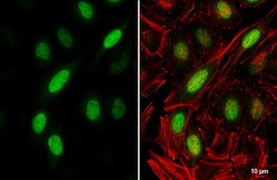
- Immunocytochemistry-Immunofluorescence analysis of Cyclin A2 was performed in HeLa cells fixed in 4% paraformaldehyde at RT for 15 min. Green: Cyclin A2 Polyclonal Antibody (Product # PA5 34682) diluted at 1:500. Red: phalloidin, a cytoskeleton marker. Scale bar = 10 µm.

- Submitted by
- Invitrogen Antibodies (provider)
- Main image
- Experimental details
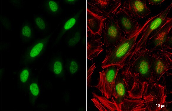
- Immunocytochemistry-Immunofluorescence analysis of Cyclin A2 was performed in HeLa cells fixed in 4% paraformaldehyde at RT for 15 min. Green: Cyclin A2 Polyclonal Antibody (Product # PA5 34682) diluted at 1:500. Red: phalloidin, a cytoskeleton marker. Scale bar = 10 µm.
Supportive validation
- Submitted by
- Invitrogen Antibodies (provider)
- Main image
- Experimental details
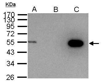
- Cyclin A2 Polyclonal Antibody immunoprecipitates cyclin A2 protein in IP experiments. IP Sample: 1,000 µg 293T whole cell lysate/extract A. 30 µg 293T whole cell lysate/extract B. Control with 2.5 µg of preimmune rabbit IgG C. Immunoprecipitation of Cyclin A2 protein by 2.5 µg of Cyclin A2 Polyclonal Antibody (Product # PA5-34682) 10% SDS-PAGE The immunoprecipitated Cyclin A2 protein was detected by Cyclin A2 Polyclonal Antibody (Product # PA5-34682) diluted at 1:1,000.

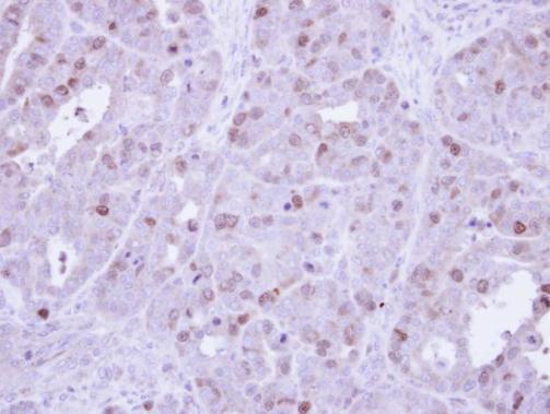
Supportive validation
- Submitted by
- Invitrogen Antibodies (provider)
- Main image
- Experimental details
- Immunohistochemical analysis of paraffin-embedded NCIN87 Xenograft, using cyclin A (Product # PA5-34682) antibody at 1:500 dilution. Antigen Retrieval: EDTA based buffer, pH 8.0, 15 min.
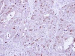
Supportive validation
- Submitted by
- Invitrogen Antibodies (provider)
- Main image

- Experimental details
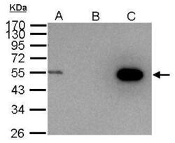
- Cyclin A2 Polyclonal Antibody immunoprecipitates cyclin A2 protein in IP experiments. IP Sample: 1,000 µg 293T whole cell lysate/extract A. 30 µg 293T whole cell lysate/extract B. Control with 2.5 µg of preimmune rabbit IgG C. Immunoprecipitation of Cyclin A2 protein by 2.5 µg of Cyclin A2 Polyclonal Antibody (Product # PA5-34682) 10% SDS-PAGE The immunoprecipitated Cyclin A2 protein was detected by Cyclin A2 Polyclonal Antibody (Product # PA5-34682) diluted at 1:1,000.