Explore
 Validate
 Learn
Learn Western blot
 Immunocytochemistry
Antibody data
- Antibody Data
- Antigen structure
- References [5]
- Comments [0]
- Validations
- Western blot [7]
- Immunocytochemistry [1]
- Immunoprecipitation [1]
- Immunohistochemistry [1]
Submit
Validation data
Reference
Comment
Report error
- Product number
- GTX101608 - Provider product page

- Provider
- GeneTex
- Proper citation
- GeneTex Cat#GTX101608, RRID:AB_1952134
- Product name
- Transgelin antibody
- Antibody type
- Polyclonal
- Reactivity
- Human, Mouse, Rat
- Host
- Rabbit
Submitted references Stellate ganglion block ameliorates vascular calcification by inhibiting endoplasmic reticulum stress.
The role of pericytic laminin in blood brain barrier integrity maintenance.
Mechanisms underlying a decrease in KCl-induced contraction after long-term serum-free organ culture of rat isolated mesenteric artery.
Biomarker discovery for neuroendocrine cervical cancer.
Depletion of the transcriptional coactivators megakaryoblastic leukaemia 1 and 2 abolishes hepatocellular carcinoma xenograft growth by inducing oncogene-induced senescence.
Hao W, Yang R, Yang Y, Jin S, Li Y, Yuan F, Guo Q, Xiao L, Wang X, Wang F, Wu Y, Teng X
Life sciences 2018 Jan 15;193:1-8
Life sciences 2018 Jan 15;193:1-8
The role of pericytic laminin in blood brain barrier integrity maintenance.
Gautam J, Zhang X, Yao Y
Scientific reports 2016 Nov 3;6:36450
Scientific reports 2016 Nov 3;6:36450
Mechanisms underlying a decrease in KCl-induced contraction after long-term serum-free organ culture of rat isolated mesenteric artery.
Morita T, Okada M, Yamawaki H
The Journal of veterinary medical science 2014 Jul;76(7):963-9
The Journal of veterinary medical science 2014 Jul;76(7):963-9
Biomarker discovery for neuroendocrine cervical cancer.
Lin LH, Chang SJ, Hu RY, Lin MW, Lin ST, Huang SH, Lyu PC, Chou HC, Lai ZY, Chuang YJ, Chan HL
Electrophoresis 2014 Jul;35(14):2039-45
Electrophoresis 2014 Jul;35(14):2039-45
Depletion of the transcriptional coactivators megakaryoblastic leukaemia 1 and 2 abolishes hepatocellular carcinoma xenograft growth by inducing oncogene-induced senescence.
Hampl V, Martin C, Aigner A, Hoebel S, Singer S, Frank N, Sarikas A, Ebert O, Prywes R, Gudermann T, Muehlich S
EMBO molecular medicine 2013 Sep;5(9):1367-82
EMBO molecular medicine 2013 Sep;5(9):1367-82
No comments: Submit comment
Enhanced validation
Supportive validation
- Submitted by
- GeneTex (provider)
- Enhanced method
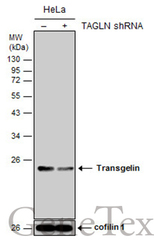
- Genetic validation
- Main image
- Experimental details
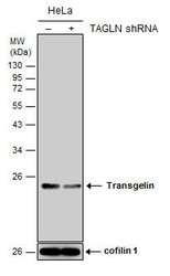
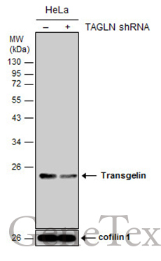
- Non-transfected (¡V) and transfected (+) HeLa whole cell extracts (15 ?g) were separated by 12% SDS-PAGE, and the membrane was blotted with Transgelin antibody (GTX101608) diluted at 1:5000. The HRP-conjugated anti-rabbit IgG antibody (GTX213110-01) was used to detect the primary antibody.
Supportive validation
- Submitted by
- GeneTex (provider)
- Main image
- Experimental details
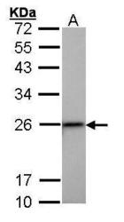
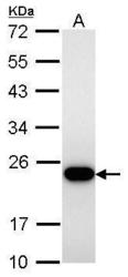
- Sample (30 ?g of whole cell lysate) A: HepG2 (GTX27900) 12% SDS PAGE GTX101608 diluted at 1:1000 The HRP-conjugated anti-rabbit IgG antibody (GTX213110-01) was used to detect the primary antibody.

- Submitted by
- GeneTex (provider)
- Main image
- Experimental details
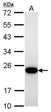
- Transgelin antibody detects TAGLN protein by western blot analysis.A. 30 ?g C8D30 whole cell lysate/extract12% SDS-PAGETransgelin antibody (GTX101608) dilution: 1:1000 The HRP-conjugated anti-rabbit IgG antibody (GTX213110-01) was used to detect the primary antibody.
- Submitted by
- GeneTex (provider)
- Main image

- Experimental details
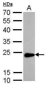
- Transgelin antibody detects TAGLN protein by western blot analysis.A. 30 ?g Rat-2 whole cell lysate/extract12% SDS-PAGETransgelin antibody (GTX101608) dilution: 1:5000 The HRP-conjugated anti-rabbit IgG antibody (GTX213110-01) was used to detect the primary antibody.
- Submitted by
- GeneTex (provider)
- Main image
- Experimental details
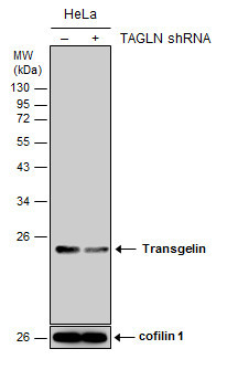
- Non-transfected (¡V) and transfected (+) HeLa whole cell extracts (15 ?g) were separated by 12% SDS-PAGE, and the membrane was blotted with Transgelin antibody (GTX101608) diluted at 1:5000. The HRP-conjugated anti-rabbit IgG antibody (GTX213110-01) was used to detect the primary antibody.
- Submitted by
- GeneTex (provider)
- Main image
- Experimental details
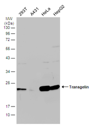
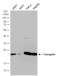
- Various whole cell extracts (30 ?g) were separated by 12% SDS-PAGE, and the membrane was blotted with Transgelin antibody (GTX101608) diluted at 1:1000. The HRP-conjugated anti-rabbit IgG antibody (GTX213110-01) was used to detect the primary antibody.
- Submitted by
- GeneTex (provider)
- Main image
- Experimental details
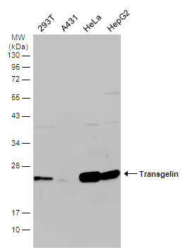
- Various whole cell extracts (30 ?g) were separated by 12% SDS-PAGE, and the membrane was blotted with Transgelin antibody (GTX101608) diluted at 1:1000. The HRP-conjugated anti-rabbit IgG antibody (GTX213110-01) was used to detect the primary antibody.

Supportive validation
- Submitted by
- GeneTex (provider)
- Main image

- Experimental details
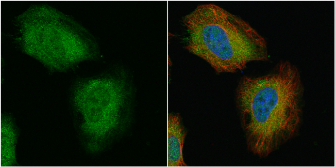
- Transgelin antibody detects Transgelin protein at cytoplasm and nucleus by immunofluorescent analysis.Sample: HeLa cells were fixed in 4% paraformaldehyde at RT for 15 min.Green: Transgelin protein stained by Transgelin antibody (GTX101608) diluted at 1:500.Red: alpha Tubulin, a cytoskeleton marker, stained by alpha Tubulin antibody [GT114] (GTX628802) diluted at 1:1000.Blue: Hoechst 33342 staining.
Supportive validation
- Submitted by
- GeneTex (provider)
- Main image
- Experimental details
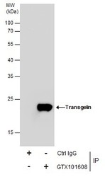
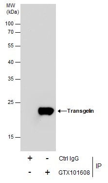
- Immunoprecipitation of Transgelin protein from HeLa whole cell extracts using 5 £gg of Transgelin antibody (GTX101608).Western blot analysis was performed using Transgelin antibody (GTX101608).EasyBlot anti-Rabbit IgG (GTX221666-01) was used as a secondary reagent.
Supportive validation
- Submitted by
- GeneTex (provider)
- Main image
- Experimental details
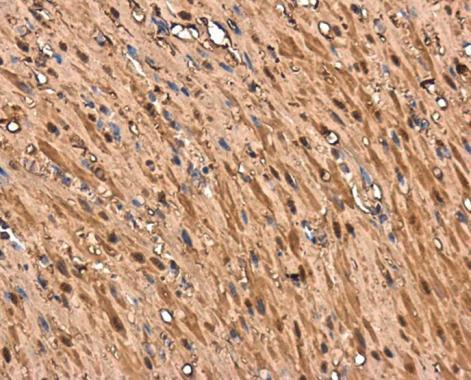
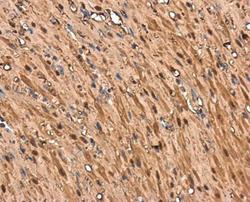
- Transgelin antibody detects Transgelin protein at cytoplasm in rat stomach by immunohistochemical analysis. Sample: Paraffin-embedded rat stomach. Transgelin antibody (GTX101608) diluted at 1:500.