Explore
 Validate
 Learn
Learn Western blot
Antibody data
- Antibody Data
- Antigen structure
- References [1]
- Comments [0]
- Validations
- Western blot [1]
- Immunohistochemistry [1]
- Flow cytometry [2]
- Other assay [1]
Submit
Validation data
Reference
Comment
Report error
- Product number
- PA5-78865 - Provider product page

- Provider
- Invitrogen Antibodies
- Product name
- Bcl-W Polyclonal Antibody
- Antibody type
- Polyclonal
- Antigen
- Synthetic peptide
- Description
- Reconstitute with 0.2 mL of distilled water to yield a concentration of 500 µg/mL. Positive Control - WB: human A549 whole cell, human K562 whole cell, human U-937 whole cell, human Hela whole cell, human A431 whole cell. IHC: human rectal cancer tissue. Flow: PC-3 cell.
- Reactivity
- Human, Mouse, Rat
- Host
- Rabbit
- Isotype
- IgG
- Vial size
- 100 μg
- Concentration
- 500 μg/mL
- Storage
- -20°C
Submitted references MicroRNA-325-3p Facilitates Immune Escape of Mycobacterium tuberculosis through Targeting LNX1 via NEK6 Accumulation to Promote Anti-Apoptotic STAT3 Signaling.
Fu B, Xue W, Zhang H, Zhang R, Feldman K, Zhao Q, Zhang S, Shi L, Pavani KC, Nian W, Lin X, Wu H
mBio 2020 Jun 2;11(3)
mBio 2020 Jun 2;11(3)
No comments: Submit comment
Supportive validation
- Submitted by
- Invitrogen Antibodies (provider)
- Main image

- Experimental details
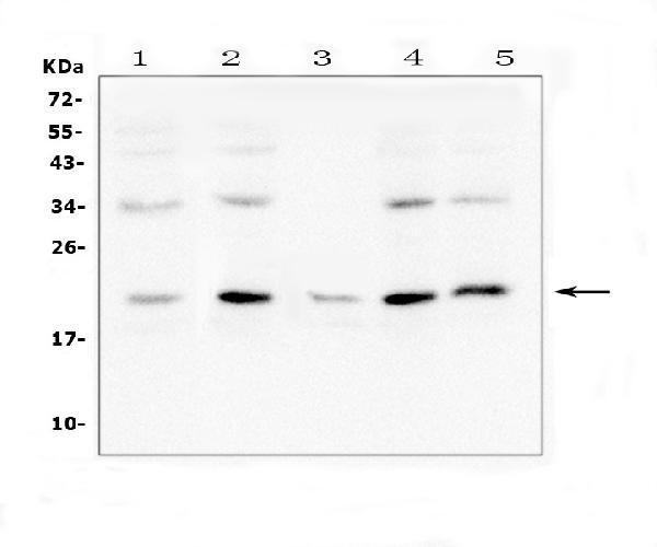
- Western blot analysis of Bcl2L2 in, Lane 1: human A549 whole cell lysates, Lane 2: human K562 whole cell lysates, Lane 3: human U-937 whole cell lysates, Lane 4: human Hela whole cell lysates., Lane 5: human A431 whole cell lysates. Electrophoresis was performed on a 5-20% SDS-PAGE gel at 70V (Stacking gel) / 90V (Resolving gel) for 2-3 hours. The sample well of each lane was loaded with 50 µg of sample under reducing conditions. After Electrophoresis, proteins were transferred to a Nitrocellulose membrane at 150mA for 50-90 minutes. The membrane was blocked with 5% Non-fat Milk/ TBS for 1.5 hour at RT. The membrane was incubated with Bcl-W Polyclonal Antibody (Product # PA5-78865) at 0.5 μg/mL overnight at 4°C, then washed with TBS-0.1%Tween 3 times with 5 minutes each and probed with a goat anti-rabbit IgG-HRP secondary antibody at a dilution of 1:10000 for 1.5 hour at RT. The signal is developed using an Enhanced Chemiluminescent detection (ECL) kit. A specific band was detected for Bcl2L2 at approximately 21KD. The expected band size for Bcl2L2 is at 21KD.
Supportive validation
- Submitted by
- Invitrogen Antibodies (provider)
- Main image
- Experimental details
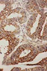
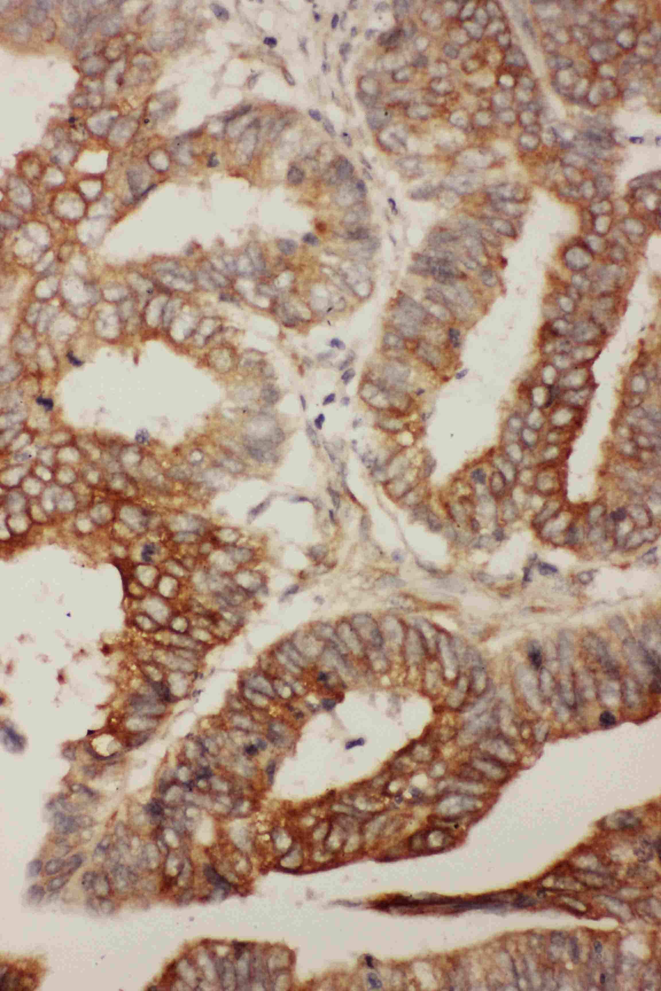
- Immunohistochemistry analysis of Bcl-W on paraffin-embedded human rectal cancer tissue. Antigen retrieval was performed using citrate buffer (pH6, epitope retrieval solution) for 20 mins. Sample was blocked using 10% goat serum, incubated with Bcl-W polyclonal antibody (Product# PA5-78865) with a dilution of 1 µg/mL (overnight at 4°C), and followed by biotinylated goat anti-rabbit IgG (30 minutes at 37°C). Development was performed using Streptavidin-Biotin-Complex (SABC) with DAB chromogen method.
Supportive validation
- Submitted by
- Invitrogen Antibodies (provider)
- Main image
- Experimental details
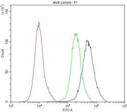
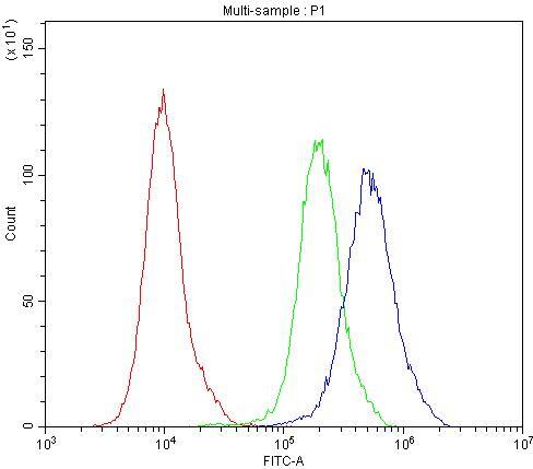
- Flow Cytometry of Bcl-W in PC-3 cells (blue line), isotype control rabbit IgG (green line) and unlabeled (red line). Samples were blocked with 10% goat serum, incubated with Bcl-W Polyclonal Antibody (Product # PA5-78865) at a dilution of 1 μg (per 1x10^6 cells), followed by DyLight®488 conjugated goat anti-rabbit IgG (for 30 minutes at 20°C) using 5-10 μg (per 1x10^6 cells) dilution.

- Submitted by
- Invitrogen Antibodies (provider)
- Main image

- Experimental details
- Flow Cytometry of Bcl-W in PC-3 cells (blue line), isotype control rabbit IgG (green line) and unlabeled (red line). Samples were blocked with 10% goat serum, incubated with Bcl-W Polyclonal Antibody (Product # PA5-78865) at a dilution of 1 μg (per 1x10^6 cells), followed by DyLight®488 conjugated goat anti-rabbit IgG (for 30 minutes at 20°C) using 5-10 μg (per 1x10^6 cells) dilution.
Supportive validation
- Submitted by
- Invitrogen Antibodies (provider)
- Main image
- Experimental details
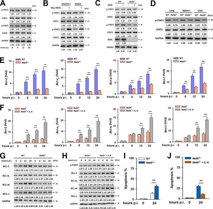
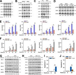
- FIG 5 NEK6 inhibits apoptosis through the activation of STAT3. (A) Immunoblot analysis of the phosphorylated STAT1 (p-STAT1), STAT1, phosphorylated STAT3 (p-STAT3), and STAT3 in RAW 264.7 cells stimulated with 10 mug/mL of gamma-irradiated M. tuberculosis . (B) Immunoblot analysis of the p-STAT1, STAT1, p-STAT3, and STAT3 in RAW 264.7 cells and BMDMs transfected with Myc-NEK6. (C) BMDMs from WT and NEK6-deficient ( Nek6 -/- ) mice were stimulated with 10 mug/mL of gamma-irradiated M. tuberculosis for 24 h. The expressions of p-STAT1, STAT1, p-STAT3, STAT3, and NEK6 were analyzed by Western blotting. (D) Immunoblot analysis of the p-STAT3 and STAT3 in lungs, spleens, and livers from WT and Nek6 -/- mice at 7 dpi. (E and G) The expression levels of BCL-2, BCL-X L , BCL-W, and MCL-1 in 10 mug/mL of gamma-irradiated M. tuberculosis -stimulated BMDMs from WT and Nek6 -/- mice were calculated by qRT-PCR at the indicated times (E) and by Western blotting (G). (F and H) BMDMs from Nek6 -/- mice were pretreated with 20 ng/mL IL-6 for 4 h h, then cells were stimulated with 10 mug/mL of gamma-irradiated M. tuberculosis . The expression levels of BCL-2, BCL-X L , BCL-W, and MCL-1 were calculated by qRT-PCR (F) and Western blotting (H) at the indicated times. (I) BMDMs from WT and Nek6 -/- mice were stimulated with 10 mug/mL of gamma-irradiated M. tuberculosis for 24 h, and the apoptosis rates were detected by flow cytometry. (J) BMDMs from Nek6 -/- mice were pretreated with 20 ng/mL of I