Explore
 Validate
 Learn
Learn Western blot
 Immunocytochemistry
Antibody data
- Antibody Data
- Antigen structure
- References [1]
- Comments [0]
- Validations
- Western blot [2]
- Immunohistochemistry [1]
Submit
Validation data
Reference
Comment
Report error
- Product number
- MAB4148 - Provider product page

- Provider
- Novus Biologicals
- Product name
- Mouse Monoclonal HSPA8/HSC71/Hsc70 Antibody
- Antibody type
- Monoclonal
- Description
- Protein A or G purified from hybridoma culture supernatant. Detects human, mouse and rat HSPA8 in Western blots.
- Reactivity
- Human, Mouse, Rat
- Host
- Mouse
- Conjugate
- Unconjugated
- Isotype
- IgG
- Vial size
- 100 ug
- Concentration
- LYOPH
- Storage
- Use a manual defrost freezer and avoid repeated freeze-thaw cycles. 12 months from date of receipt, -20 to -70 degreesC as supplied. 1 month, 2 to 8 degreesC under sterile conditions after reconstitution. 6 months, -20 to -70 degreesC under sterile conditions after reconstitution.
Submitted references Angiopoietin-like protein 2 accelerates carcinogenesis by activating chronic inflammation and oxidative stress.
Aoi J, Endo M, Kadomatsu T, Miyata K, Ogata A, Horiguchi H, Odagiri H, Masuda T, Fukushima S, Jinnin M, Hirakawa S, Sawa T, Akaike T, Ihn H, Oike Y
Molecular cancer research : MCR 2014 Feb;12(2):239-49
Molecular cancer research : MCR 2014 Feb;12(2):239-49
No comments: Submit comment
Supportive validation
- Submitted by
- Novus Biologicals (provider)
- Main image
- Experimental details
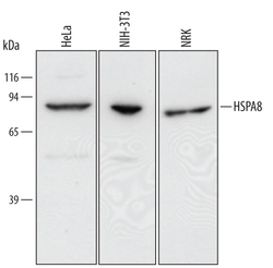
- Detection of Human/Mouse/Rat HSPA8/HSC71 by Western Blot. Western blot shows lysates of HeLa human cervical epithelial carcinoma cell line, NIH-3T3 mouse embryonic fibroblast cell line, and NRK rat normal kidney cell line. PVDF membrane was probed with 0.1 µg/mL of Mouse Anti-Human/Mouse/Rat HSPA8/HSC71 Monoclonal Antibody (Catalog # MAB4148) followed by HRP-conjugated Anti-Mouse IgG Secondary Antibody (Catalog # HAF007). A specific band was detected for HSPA8/HSC71 at approximately 75 kDa (as indicated). This experiment was conducted under reducing conditions and using Immunoblot Buffer Group 2.
- Submitted by
- Novus Biologicals (provider)
- Main image

- Experimental details
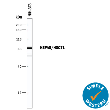
- Detection of Mouse HSPA8/HSC71 by Simple WesternTM. Simple Western lane view shows lysates of NIH-3T3 mouse embryonic fibroblast cell line, loaded at 0.2 mg/mL. A specific band was detected for HSPA8/HSC71 at approximately 93 kDa (as indicated) using 1 µg/mL of Mouse Anti-Human/Mouse/Rat HSPA8/HSC71 Monoclonal Antibody (Catalog # MAB4148). This experiment was conducted under reducing conditions and using the 12-230 kDa separation system.
Supportive validation
- Submitted by
- Novus Biologicals (provider)
- Main image
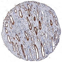
- Experimental details
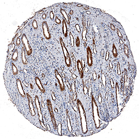
- HSPA8/HSC71 in Human Kidney. HSPA8/HSC71 was detected in immersion fixed paraffin-embedded sections of human kidney using Mouse Anti-Human/Mouse/Rat HSPA8/HSC71 Monoclonal Antibody (Catalog # MAB4148) at 5 µg/mL for 1 hour at room temperature followed by incubation with the Anti-Mouse IgG VisUCyte™ HRP Polymer Antibody (Catalog # VC001). Tissue was stained using DAB (brown) and counterstained with hematoxylin (blue). Specific staining was localized to epithelial cells in convoluted tubules. View our protocol for IHC Staining with VisUCyte HRP Polymer Detection Reagents.