 Explore
 Validate
 Learn
Learn Western blot
 Immunocytochemistry
Antibody data
- Antibody Data
- Antigen structure
- References [2]
- Comments [0]
- Validations
- Immunocytochemistry [2]
- Immunohistochemistry [1]
- Flow cytometry [2]
- Other assay [2]
Submit
Validation data
Reference
Comment
Report error
- Product number
- PA5-24624 - Provider product page

- Provider
- Invitrogen Antibodies
- Product name
- HSC70 Polyclonal Antibody
- Antibody type
- Polyclonal
- Antigen
- Synthetic peptide
- Description
- This antibody is predicted to react with bovine, chicken, hamster, mouse and rat based on sequence homology.
- Reactivity
- Human, Mouse, Rat
- Host
- Rabbit
- Isotype
- IgG
- Vial size
- 400 μL
- Concentration
- 1.5 mg/mL
- Storage
- Store at 4°C short term. For long term storage, store at -20°C, avoiding freeze/thaw cycles.
Submitted references A Proteomic Study Suggests Stress Granules as New Potential Actors in Radiation-Induced Bystander Effects.
Multi-faceted proteomic characterization of host protein complement of Rift Valley fever virus virions and identification of specific heat shock proteins, including HSP90, as important viral host factors.
Tudor M, Gilbert A, Lepleux C, Temelie M, Hem S, Armengaud J, Brotin E, Haghdoost S, Savu D, Chevalier F
International journal of molecular sciences 2021 Jul 26;22(15)
International journal of molecular sciences 2021 Jul 26;22(15)
Multi-faceted proteomic characterization of host protein complement of Rift Valley fever virus virions and identification of specific heat shock proteins, including HSP90, as important viral host factors.
Nuss JE, Kehn-Hall K, Benedict A, Costantino J, Ward M, Peyser BD, Retterer CJ, Tressler LE, Wanner LM, McGovern HF, Zaidi A, Anthony SM, Kota KP, Bavari S, Hakami RM
PloS one 2014;9(5):e93483
PloS one 2014;9(5):e93483
No comments: Submit comment
Supportive validation
- Submitted by
- Invitrogen Antibodies (provider)
- Main image
- Experimental details
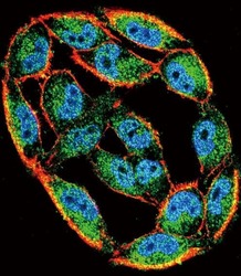
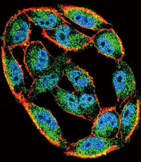
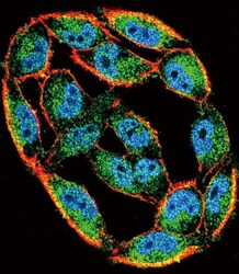
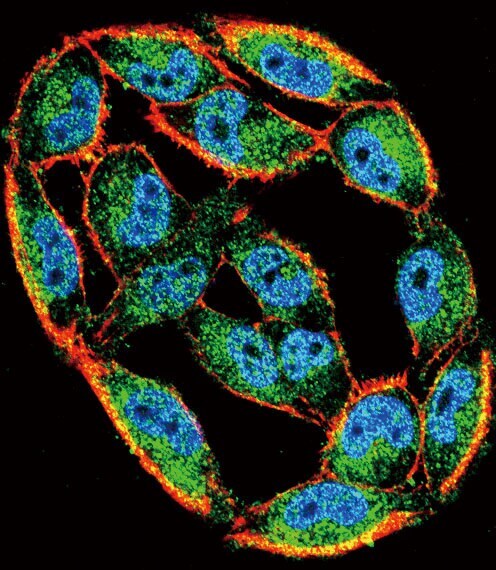
- Immunofluorescent analysis of A2058 cells using a HSPA8/Heat Shock 70 Protein 8 polyclonal antibody (Product # PA5-24624) at a dilution of 1:10-50, followed by a fluor-conjugated goat anti-rabbit secondary antibody (green). Actin filaments were stained with dye-conjugated phalloidin (red). Nuclei were stained with DAPI (blue).
- Submitted by
- Invitrogen Antibodies (provider)
- Main image
- Experimental details
- Immunocytochemistry analysis of HSC70 in A2058 cells. Samples were incubated in HSC70 polyclonal antibody (Product # PA5-24624) followed by Alexa Fluor 488-conjugated goat anti-rabbit lgG (green). Actin filaments have been labeled with Alexa Fluor 555 phalloidin (red). DAPI was used to stain the cell nuclear (blue).
Supportive validation
- Submitted by
- Invitrogen Antibodies (provider)
- Main image
- Experimental details
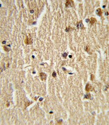
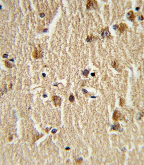
- Immunohistochemistry analysis of HSC70 in formalin-fixed and paraffin-embedded human brain tissue. Samples were incubated with HSC70 polyclonal antibody (Product # PA5-24624) which was peroxidase-conjugated to the secondary antibody, followed by DAB staining. This data demonstrates the use of this antibody for immunohistochemistry; clinical relevance has not been evaluated.
Supportive validation
- Submitted by
- Invitrogen Antibodies (provider)
- Main image
- Experimental details
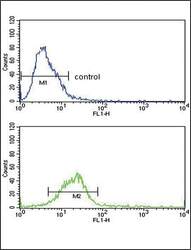
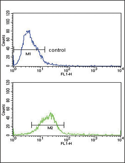
- Flow cytometry analysis of HeLa cells using a HSPA8/Heat Shock 70 Protein 8 polyclonal antibody (Product # PA5-24624) (bottom) compared to a negative control cell (top) at a dilution of 1:10-50, followed by a FITC-conjugated goat anti-rabbit antibody
- Submitted by
- Invitrogen Antibodies (provider)
- Main image

- Experimental details
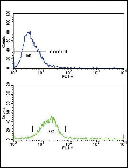
- Flow cytometry of HSC70 in Hela cells (bottom histogram). Samples were incubated with HSC70 polyclonal antibody (Product # PA5-24624) followed by FITC-conjugated goat-anti-rabbit secondary antibody. Negative control cell (top histogram).
Supportive validation
- Submitted by
- Invitrogen Antibodies (provider)
- Main image
- Experimental details
- NULL
- Submitted by
- Invitrogen Antibodies (provider)
- Main image
- Experimental details
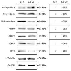
- Figure 5 Western blotting analysis of cyclophilin A, thioredoxin, alpha-enolase, RPLP0, HSC70, HSPA9, and CCT3 in a whole-cell extracts from T/C-28A2 bystander cells receiving the conditioned medium of low-dose irradiated chondrosarcoma cells (0.1 Gy) or non-irradiated chondrosarcoma cells (CTR).
