 Explore
 Validate
 Learn
Learn Western blot
Antibody data
- Antibody Data
- Antigen structure
- References [1]
- Comments [0]
- Validations
- Western blot [5]
- Immunocytochemistry [1]
- Immunohistochemistry [3]
- Other assay [1]
Submit
Validation data
Reference
Comment
Report error
- Product number
- PA5-29221 - Provider product page

- Provider
- Invitrogen Antibodies
- Product name
- HSC70 Polyclonal Antibody
- Antibody type
- Polyclonal
- Antigen
- Recombinant protein fragment
- Description
- Recommended positive controls: A431, HeLa, Neuro2A, GL261, C8D30, 293T, HepG2. Predicted reactivity: Mouse (100%), Rat (100%), Zebrafish (97%), Xenopus laevis (97%), Pig (100%), Chicken (98%), Rhesus Monkey (100%), Bovine (100%). Store product as a concentrated solution. Centrifuge briefly prior to opening the vial.
- Reactivity
- Human, Mouse
- Host
- Rabbit
- Isotype
- IgG
- Vial size
- 100 µL
- Concentration
- 0.25 mg/mL
- Storage
- Store at 4°C short term. For long term storage, store at -20°C, avoiding freeze/thaw cycles.
Submitted references Deletion of translin (Tsn) induces robust adiposity and hepatic steatosis without impairing glucose tolerance.
Shah AP, Johnson MD, Fu X, Boersma GJ, Shah M, Wolfgang MJ, Tamashiro KL, Baraban JM
International journal of obesity (2005) 2020 Jan;44(1):254-266
International journal of obesity (2005) 2020 Jan;44(1):254-266
No comments: Submit comment
Supportive validation
- Submitted by
- Invitrogen Antibodies (provider)
- Main image
- Experimental details
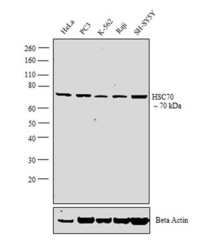
- Western blot analysis was performed on whole cell extracts (30 µg lysate) of HeLa (Lane 1), PC3 (Lane 2), K-562 (Lane 3), Raji (Lane 4) and SH-SY5Y (Lane 5). The blot was probed with Anti-HSC70 Polyclonal Antibody (Product # PA5-29221, 1:1000 dilution) and detected by chemiluminescence using Goat anti-Rabbit IgG (H+L) Superclonal™ Secondary Antibody, HRP conjugate (Product # A27036, 0.25 µg/ml, 1:4000 dilution). A 70 kDa band corresponding to HSC70 was observed across all the cell lines tested.

- Submitted by
- Invitrogen Antibodies (provider)
- Main image
- Experimental details
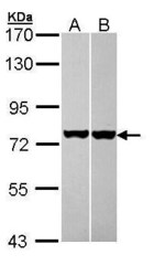
- Western Blot using HSC70 Polyclonal Antibody (Product # PA5-29221). Sample (30 µg of whole cell lysate). Lane A: A431. Lane B: HeLa. 7.5% SDS PAGE. HSC70 Polyclonal Antibody (Product # PA5-29221) diluted at 1:1,000. The HRP-conjugated anti-rabbit IgG antibody was used to detect the primary antibody.

- Submitted by
- Invitrogen Antibodies (provider)
- Main image
- Experimental details
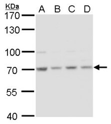
- HSC70 Polyclonal Antibody detects HSC70 protein by western blot analysis. A. 30 µg 293T whole cell lysate/extract. B. 30 µg A431whole cell lysate/extract. C. 30 µg HeLa whole cell lysate/extract. D. 30 µg HepG2 whole cell lysate/extract.7.5% SDS-PAGE. HSC70 Polyclonal Antibody (Product # PA5-29221) dilution: 1:1,000. The HRP-conjugated anti-rabbit IgG antibody was used to detect the primary antibody.

- Submitted by
- Invitrogen Antibodies (provider)
- Main image
- Experimental details
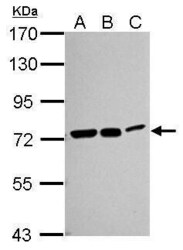
- Western Blot using HSC70 Polyclonal Antibody (Product # PA5-29221). Sample (30 µg of whole cell lysate). Lane A: Neuro2A. Lane B: GL261. Lane C: C8D30. 7.5% SDS PAGE. HSC70 Polyclonal Antibody (Product # PA5-29221) diluted at 1:1,000. The HRP-conjugated anti-rabbit IgG antibody was used to detect the primary antibody.

- Submitted by
- Invitrogen Antibodies (provider)
- Main image
- Experimental details
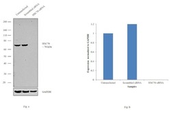
- Knockdown of HSC70 was achieved by transfecting HeLa with HSC70 specific siRNAs (Silencer® select Product # s6987). Western blot analysis (Fig. a) was performed using whole cell extracts from the HSC70 knockdown cells (lane 3), non-specific scrambled siRNA transfected cells (lane 2) and untransfected cells (lane 1). The blot was probed with HSC70 Polyclonal Antibody (Product # PA5-29221, 1:2000 dilution) and Goat anti-Rabbit IgG (H+L) Superclonal™ Secondary Antibody, HRP conjugate (Product # A27036, 0.25µg/ml, 1:4000 dilution). Densitometric analysis of this western blot is shown in histogram (Fig. b). Decrease in signal upon siRNA mediated knock down confirms that antibody is specific to HSC70.

Supportive validation
- Submitted by
- Invitrogen Antibodies (provider)
- Main image

- Experimental details
- HSC70 Polyclonal Antibody detects HSC70 protein at cytoplasm by immunofluorescent analysis. Sample: HeLa cells were fixed in ice-cold MeOH for 5 min. Green: HSC70 protein stained by HSC70 Polyclonal Antibody (Product # PA5-29221) diluted at 1:500. Blue: Hoechst 33342 staining.

Supportive validation
- Submitted by
- Invitrogen Antibodies (provider)
- Main image
- Experimental details
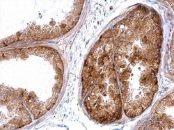
- HSC70 antibody detects HSC70 protein at cytosol on mouse prostate by immunohistochemical analysis. Sample: Paraffin-embedded mouse prostate. HSC70 antibody (Product # PA5-29221) dilution: 1:500. Antigen Retrieval: EDTA based buffer, pH 8.0, 15 min.

- Submitted by
- Invitrogen Antibodies (provider)
- Main image
- Experimental details
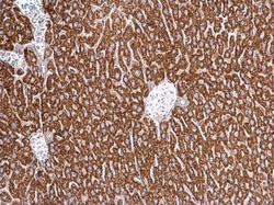
- HSC70 antibody detects HSC70 protein at cytosol on mouse prostate by immunohistochemical analysis. Sample: Paraffin-embedded mouse prostate. HSC70 antibody (Product # PA5-29221) dilution: 1:500. Antigen Retrieval: EDTA based buffer, pH 8.0, 15 min.
- Submitted by
- Invitrogen Antibodies (provider)
- Main image

- Experimental details
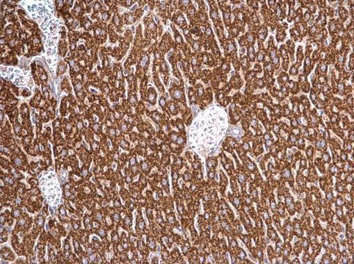
- Immunohistochemical analysis of paraffin-embedded NCIN87 xenograft, using HSC70 (Product # PA5-29221) antibody at 1:100 dilution. Antigen Retrieval: EDTA based buffer, pH 8.0, 15 min.
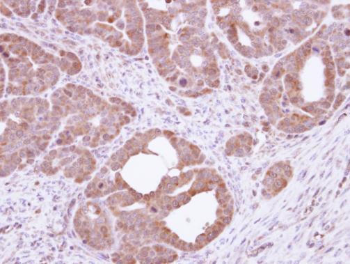
Supportive validation
- Submitted by
- Invitrogen Antibodies (provider)
- Main image

- Experimental details
- Figure 5: Characterization of adipose tissue. A: Representative images of H&E stained eWAT. Scale bars, 100mu. B: Percentage frequency distribution of adipocyte area (left), Friedman test showed a significant difference between the distributions ( P = 0.0042) and correlation between %fat mass and average adipocyte area (in mum 2 ) for WT-LFD ( r = 0.95, P = 0.0139), WT-HFD ( r = 0.85, P = 0.0317) and KO-LFD ( r = 0.83, P = 0.0415) (right). C: Representative bands for trax and translin protein in eWAT (left) and their quantification normalized to heat shock cognate 71 kDa (Hsc70) (right). n = 5, 6 and 6 for the three groups, respectively. Data are expressed as Mean +- SEM. Statistical significance was assessed by Student's t -test, ordinary or Friedman's test followed by Dunn's post-hoc analysis; (white circles/bars = WT-LFD; grey squares/bars = WT-HFD; black triangles = KO-LFD).
