 Explore
 Validate
 Learn
LearnPA5-29042
antibody from Invitrogen Antibodies
Targeting: HSP90AA1
FLJ31884, Hsp89, Hsp90, HSP90N, HSPC1, HSPCA
 Western blot
Antibody data
- Antibody Data
- Antigen structure
- References [0]
- Comments [0]
- Validations
- Western blot [5]
- Immunocytochemistry [2]
- Immunohistochemistry [6]
Submit
Validation data
Reference
Comment
Report error
- Product number
- PA5-29042 - Provider product page

- Provider
- Invitrogen Antibodies
- Product name
- HSP90 alpha Polyclonal Antibody
- Antibody type
- Polyclonal
- Antigen
- Recombinant protein fragment
- Description
- Recommended positive controls: 293T, A431, HeLa, HepG2, mouse brain, rat brain, RK3E.
- Concentration
- 0.27 mg/mL
No comments: Submit comment
Supportive validation
- Submitted by
- Invitrogen Antibodies (provider)
- Main image

- Experimental details
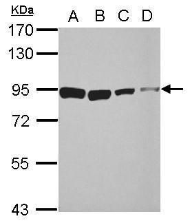
- Western blot analysis of HSP90/Heat Shock Protein 90 alpha using 30 µg of A) 293T (B) A431 (C) HeLa and D) HepG2 lysate. Samples were loaded onto a 7.5% SDS-PAGE gel and probed with a HSP90/Heat Shock Protein 90 alpha polyclonal antibody (Product # PA5-29042) at a dilution of 1:10,000.
- Submitted by
- Invitrogen Antibodies (provider)
- Main image

- Experimental details
- Western blot analysis was performed on whole cell extracts (30 µg lysate) of A-431 (Lane 1), U-87 MG (Lane 2), HEK 293T (Lane 3), HeLa (Lane 4), K-562 (Lane 5) and NIH/3T3 (Lane 6). The blot was probed with Anti-HSP90 alpha Polyclonal Antibody (Product # PA5-29042, 1 µg/mL dilution) and detected by chemiluminescence using Goat anti-Rabbit IgG (H+L) Superclonal™ Secondary Antibody, HRP conjugate (Product # A27036, 0.25 µg/mL, 1:4000 dilution). A 80 kDa band corresponding to HSP90 alpha was observed across the cell lines tested.

- Submitted by
- Invitrogen Antibodies (provider)
- Main image
- Experimental details
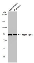
- Western Blot analysis of HSP90 alpha was performed by separating 50 µg of Various tissue extracts by 7.5% SDS-PAGE. Proteins were transferred to a membrane and probed with a HSP90 alpha Polyclonal Antibody (Product # PA5-29042) at a dilution of 1:10000. The HRP-conjugated anti-rabbit IgG antibody was used to detect the primary antibody.

- Submitted by
- Invitrogen Antibodies (provider)
- Main image

- Experimental details
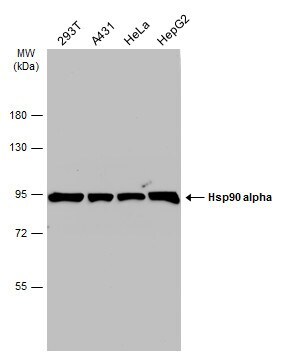
- Western Blot analysis of HSP90 alpha was performed by separating 30 µg of various whole cell extracts by 7.5% SDS-PAGE. Proteins were transferred to a membrane and probed with a HSP90 alpha Polyclonal Antibody (Product # PA5-29042) at a dilution of 1:10000 and a HRP-conjugated anti-rabbit IgG secondary antibody.
- Submitted by
- Invitrogen Antibodies (provider)
- Main image

- Experimental details
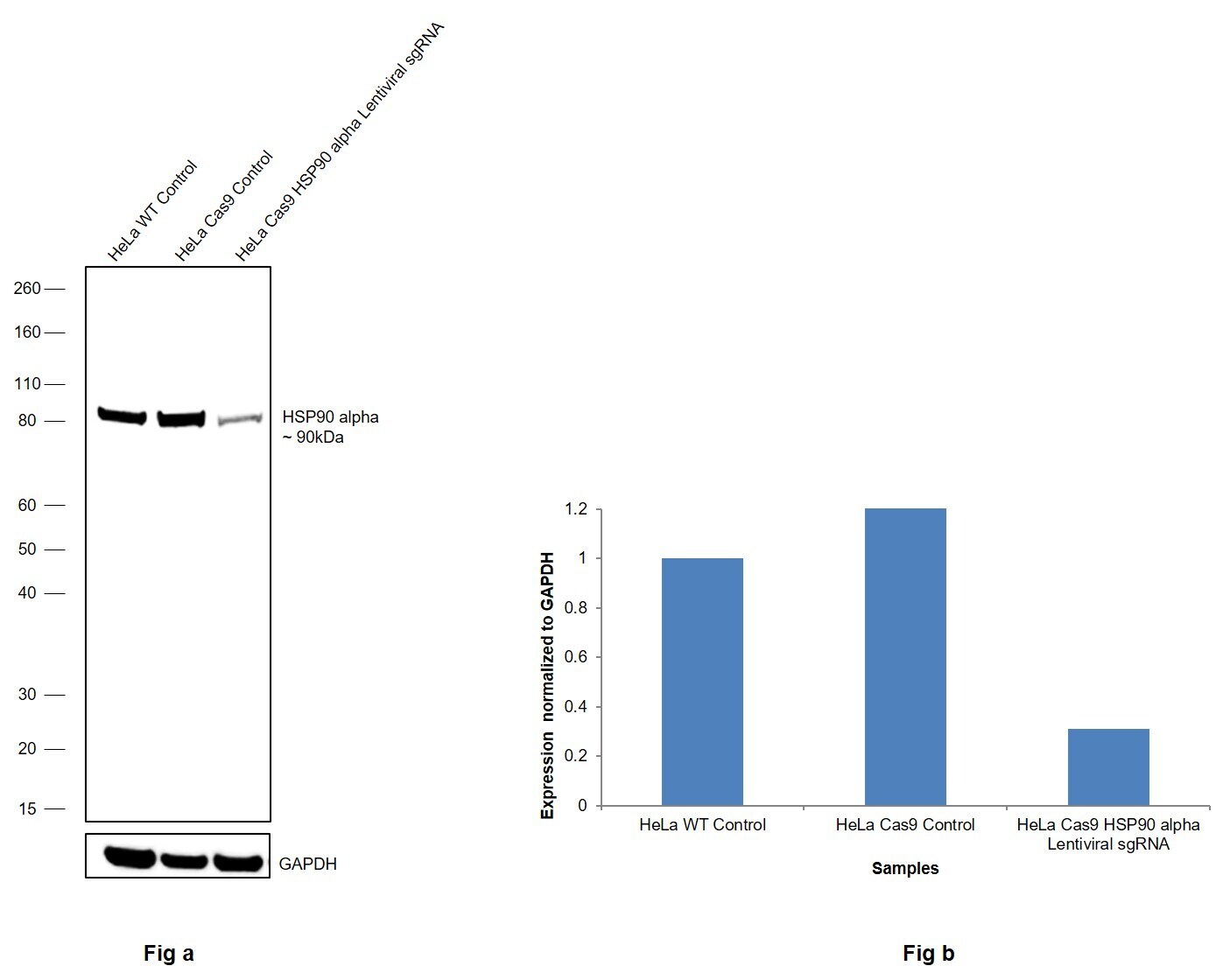
- CRISPR-Cas9 mediated genome editing ofHSP90 alpha (as confirmed by next generation sequencing) was achieved by using LentiArray™ Lentiviral sgRNA (Product # A32042, AssayID CRISPR610567_LV) and LentiArray Cas9 Lentivirus (Product # A32064). Fig (a) Western blot analysis of HSP90 alpha was performed by loading 30 µg of HeLa Wild Type (Lane 1), HeLa Cas9 (Lane 2) and HeLa Cas9 cells transduced with HSP90 alpha Lentiviral sgRNA (Lane 3) whole cell extracts. The samples were electrophoresed using NuPAGE™ Novex™ 4-12% Bis-Tris Protein Gel (Product # NP0322BOX). Resolved proteins were then transferred onto a nitrocellulose membrane (Product # IB23001) by iBlot® 2 Dry Blotting System (Product # IB21001). The blot was probed with Anti-HSP90 alpha Polyclonal Antibody (Product # PA5-29042) using 1:2,000 dilution and Goat anti-Rabbit IgG (H+L) Superclonal™ Recombinant Secondary Antibody, HRP (Product # A27036 1:8,000 dilution).Chemiluminescent detection was performed using Novex® ECL Chemiluminescent Substrate Reagent Kit (Product # WP20005). A reduced signal in sgRNA transduced cells using the LentiArray™ CRISPR product line confirms that antibody is specific toHSP90 alpha (Fig (b)).
Supportive validation
- Submitted by
- Invitrogen Antibodies (provider)
- Main image

- Experimental details
- Immunofluorescence analysis of HSP90 alpha was performed using 70% confluent log phase HeLa cells. The cells were fixed with 4% paraformaldehyde for 10 minutes, permeabilized with 0.1% Triton™ X-100 for 10 minutes, and blocked with 1% BSA for 1 hour at room temperature. The cells were labeled with HSP90 alpha Polyclonal Antibody (Product # PA5-29042) at 5 µg/mL in 0.1% BSA and incubated overnight at 4 degree and then labeled with Goat anti-Rabbit IgG (H+L) Superclonal™ Secondary Antibody, Alexa Fluor® 488 conjugate (Product # A27034) at a dilution of 1:2000 for 45 minutes at room temperature (Panel a: green). Nuclei (Panel b: blue) were stained with SlowFade® Gold Antifade Mountant with DAPI (Product # S36938). F-actin (Panel c: red) was stained with Rhodamine Phalloidin (Product # R415, 1:300). Panel d represents the merged image showing cytoplasmic and nuclear localization. Panel e represents control cells with no primary antibody to assess background. The images were captured at 60X magnification.

- Submitted by
- Invitrogen Antibodies (provider)
- Main image

- Experimental details
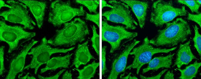
- Immunocytochemistry-Immunofluorescence analysis of HSP90 alpha was performed in HeLa cells fixed in ice cold MeOH for 5 min. Green: HSP90 alpha Polyclonal Antibody (Product # PA5 29042) diluted at 1:500. Blue: Hoechst 33342 staining.
Supportive validation
- Submitted by
- Invitrogen Antibodies (provider)
- Main image

- Experimental details
- Immunohistochemistry (Paraffin) analysis of HSP90 alpha was performed in paraffin-embedded mouse testis tissue using HSP90 alpha Polyclonal Antibody (Product # PA5-29042) at a dilution of 1:500.

- Submitted by
- Invitrogen Antibodies (provider)
- Main image

- Experimental details
- Immunohistochemistry (Paraffin) analysis of HSP90 alpha was performed in paraffin-embedded human colon carcinoma tissue using HSP90 alpha Polyclonal Antibody (Product # PA5-29042) at a dilution of 1:500.

- Submitted by
- Invitrogen Antibodies (provider)
- Main image

- Experimental details
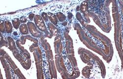
- HSP90 alpha Polyclonal Antibody detects Hsp90 alpha protein at cytoplasm by immunohistochemical analysis. Sample: Paraffin-embedded mouse duodenum. Hsp90 alpha stained by HSP90 alpha Polyclonal Antibody (Product # PA5-29042) diluted at 1:1,000. Antigen Retrieval: Citrate buffer, pH 6.0, 15 min.

- Submitted by
- Invitrogen Antibodies (provider)
- Main image
- Experimental details
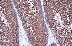
- HSP90 alpha Polyclonal Antibody detects Hsp90 alpha protein at cytoplasm by immunohistochemical analysis. Sample: Paraffin-embedded mouse intestine. Hsp90 alpha stained by HSP90 alpha Polyclonal Antibody (Product # PA5-29042) diluted at 1:1,000. Antigen Retrieval: Citrate buffer, pH 6.0, 15 min.

- Submitted by
- Invitrogen Antibodies (provider)
- Main image
- Experimental details
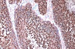
- HSP90 alpha Polyclonal Antibody detects Hsp90 alpha protein at cytoplasm by immunohistochemical analysis. Sample: Paraffin-embedded rat testis. Hsp90 alpha stained by HSP90 alpha Polyclonal Antibody (Product # PA5-29042) diluted at 1:1,000. Antigen Retrieval: Citrate buffer, pH 6.0, 15 min.

- Submitted by
- Invitrogen Antibodies (provider)
- Main image
- Experimental details
- HSP90 alpha Polyclonal Antibody detects Hsp90 alpha protein at cytoplasm by immunohistochemical analysis. Sample: Paraffin-embedded rat testis. Hsp90 alpha stained by HSP90 alpha Polyclonal Antibody (Product # PA5-29042) diluted at 1:1,000. Antigen Retrieval: Citrate buffer, pH 6.0, 15 min.
